(Left) Graphic shows a fistula between the transverse duodenum and aorta at the site of the graft-aortic suture line .
(Right) Axial CECT in a 70-year-old man presenting with fever and hematemesis months after abdominal aortic aneurysm (AAA) repair shows the native, calcified aortic wall wrapped around a synthetic graft. A gas collection is noted between the graft and aortic wall , indicating infection or fistula. Note the soft tissue density surrounding the aorta and 3rd portion of duodenum .
(Left) This elderly woman had pain and fever years after AAA repair. Axial CECT shows a calcified aortic wall wrapped around synthetic graft material . At the level of the 3rd portion of the duodenum, the duodenal wall appears to be adherent to the aorta. Note an enhanced focus that may represent active bleeding or inflammation.
(Right) Axial CECT in the same patient reveals several bubbles of extraluminal gas ; aortoenteric fistula with underlying infection was surgically confirmed.
TERMINOLOGY
Definitions
• Abnormal communication between aorta and gastrointestinal (GI) tract
IMAGING
General Features
• Best diagnostic clue
Inflammatory stranding and gas between abdominal aorta and 3rd part of duodenum following aneurysm repair
• Location
Duodenum (80%); jejunum and ileum (10-15%); stomach and colon (5%)
 between the transverse duodenum and aorta at the site of the graft-aortic suture line
  .
.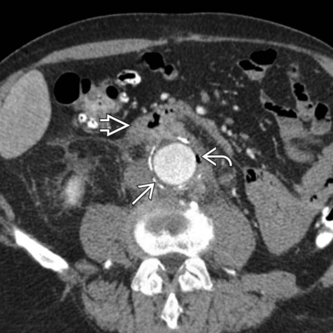
 wrapped around a synthetic graft. A gas collection is noted between the graft and aortic wall
  , indicating infection or fistula. Note the soft tissue density surrounding the aorta and 3rd portion of duodenum
  .
.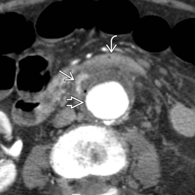
 . At the level of the 3rd portion of the duodenum, the duodenal wall
  appears to be adherent to the aorta. Note an enhanced focus
  that may represent active bleeding or inflammation.
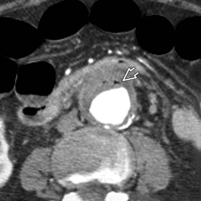
 ; aortoenteric fistula with underlying infection was surgically confirmed.


 PET/CT may be even better


