16 Appearance of Common Masses
♦Common Solid Neoplasms in the Hand and Wrist
Giant Cell Tumor of the Tendon Sheath
• A giant cell tumor of the tendon sheath is an idiopathic benign soft tissue tumor constituted of synovial-like cells including multinucleated giant cells. These masses are pathologically identical to pigmented villonodular synovitis.
• The giant cell tumor is the second most common benign mass in the hand and wrist.
○ The most common age distribution is 30 to 50 years; women are more commonly affected than men.
○ In the hand, a giant cell tumor of the tendon sheath has a predilection for the radial three digits (index and long finger), particularly around the volar aspect and distal interphalangeal joints.
○ These masses most commonly present as a chronic painless swelling or firm mass in the absence of trauma.
• Giant cell tumors have an intimate involvement with the hand and wrist tendon sheaths, because they arise from the tendon sheath synovial lining. Flexor tendon involvement is more common than extensor tendon involvement.
○ There can be a variable length and circumference of contact between the mass and the tendon sheath.
○ Masses may insinuate between the tendon and underlying bone.
○ Dynamic imaging demonstrates no excursion of the mass relative to the tendon with digit flexion or extension.
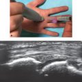
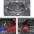
• These masses present sonographically as a predominantly homogeneously hypoechoic solid mass (Figs. 16.1 and 16.2). The presence of internal heterogeneity may be dependent on size. Additional ultrasound features may be noted:
○ Lobulated, well-demarcated margins
○ Occasional posterior acoustic enhancement
○ Absence of cystic or necrotic changes, calcification, or posterior shadowing
• Giant cell tumors of the tendon sheath typically have internal vascularity.
• A giant cell tumor of the tendon sheath is treated with surgical excision. After surgery, the recurrence rate is 10 to 20%.

Fig. 16.1 A hypoechoic soft tissue mass in the palmar second digit (arrowheads), with a neck interposed between the flexor tendons (dashed arrow) and middle phalanx (solid arrow).

Fig. 16.2 Axial T1 image of a T1 hypointense mass in the volar second digit, contacting the second digit flexor tendon and insinuating between the tendon and middle phalanx.
Lipoma
• Lipomas are benign soft tissue masses composed of mature adipocytes.
○ Throughout the body, lipomas are the most common overall soft tissue tumor. However, only approximately 5% occur in the hand or wrist.
○ A lipoma typically presents as a slow growing, mobile soft mass, usually asymptomatic.
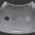
• An elongated, well-delineated morphology is typically described with the lesion parallel to the subcutaneous fat and skin surface; that is, the lesion is longer than it is wide (Fig. 16.3).
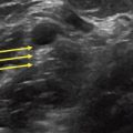
• Sonographic features of lipomas commonly include an internal echogenicity and echotexture that is similar to the surrounding fat (Fig. 16.4).
○ However, these masses may range from hypoechoic to hyperechoic with interspersed linear areas of increased echogenicity.
○ Signal characteristics are dependent on the echogenicity of surrounding structures and the degree of fat and connective tissue composition.
• Internal vascularity is typically absent.
• The presence of mass enlargement, associated pain, deep location, and irregular margins are indications for characterization with further diagnostic imaging such as MRI or CT.

Fig. 16.3 (a) A well-circumscribed mass in the subcutaneous soft tissues of the upper arm with homogeneous echotexture and that is isoechoic to the surrounding subcutaneous fat. (b) No internal hypervascularity was seen with color Doppler imaging.

Fig. 16.4 A homogeneously hyperechoic mass in the subcutaneous fat of the forearm with illdefined borders, representing a poorly encapsulated lipoma or angiomyolipoma (lipoma variant).
Hemangioma
• Hemangiomas are the most common benign vascular neoplasm, encompassing up to 7% of benign soft tissue tumors.
• Characteristic sonographic features of hemangiomas include the presence of anechoic to hypoechoic vascular channels with interspersed hyperechoic areas that occur at the interfaces between intralesional fatty tissue and surrounding neoplastic vessels (Fig. 16.5a).
• Hemangiomas often demonstrate a variable morphology, ranging from well-defined soft tissue masses to an infiltrative tangle of vessels.
• Low flow within vascular channels is commonly noted on color or power Doppler imaging (Fig. 16.5b,c).
○ Internal echogenic, shadowing foci representing phleboliths may be present.
○ Channels may demonstrate increased echogenicity if thrombosed.
○ Larger lesions may manifest compressible vascular channels with provocative maneuvers.
○ The presence of arteriovenous shunting typically indicates true arteriovenous malformation.