24 Arteries of the Female Genital Tract
T. Kroencke
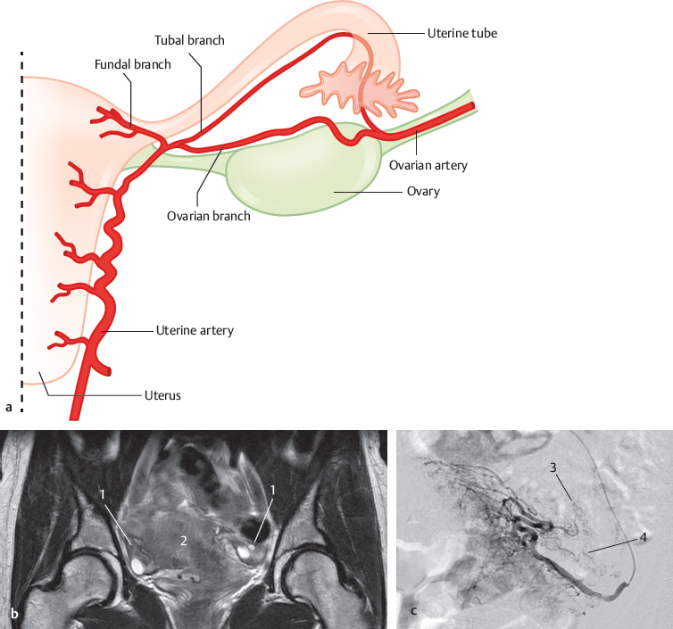
The internal female genital organs are supplied by the uterine and ovarian arteries. The origin of the ovarian artery is shown in Chapter 12 and that of the uterine artery in the chapter on the internal iliac artery (Chapter 23). Both arteries anastomose by branches along the uterine tube. The ovary seems to be supplied either by the ovarian artery or by the ovarian branch of the uterine artery, or by both. Operations on the uterine tube can disturb the blood flow in these branches. There are systematic studies on anomalies in the course and the size of the anastomosing branch between the ovarian and uterine arteries. With the increasing number of uterine artery embolization for treatment of fibroids, ovarian artery-to-uterine artery anastomoses have been studied as a potential source of treatment failure and nontarget embolization.1–11
24.1 “Normal” Situation as Described in Textbook (>90%)

Fig. 24.1 “Normal” situation as described in textbooks (>90%). Schematic (a), T2-weighted coronal MRI (b), and selective angiography of the left uterine artery (c). 1 Ovary; 2 uterus; 3 ovarian branch; 4 tubal branch.
24.2 Blood Supply of the Fundus of the Uterus

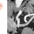
Fig. 24.2 Uterine artery (90%). Schematic (a) and selective angiography of the uterine artery (b). 1 Angiographic catheter; 2 fundal branch of the uterine artery; 3 arcuate artery.

Fig. 24.3 Branches of the ovarian artery (10%). Schematic (a) and selective angiography of the ovarian artery post uterine artery embolization (opacification of fibroid) (b). 1 Ovarian artery; 2 ovarian artery terminating as fundal branches; uterine fibroid (*).
Stay updated, free articles. Join our Telegram channel

Full access? Get Clinical Tree


