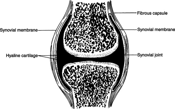
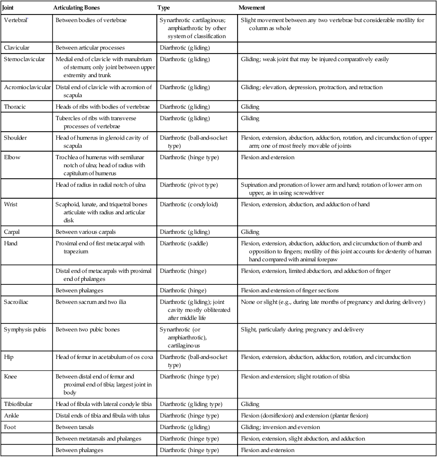
CHAPTER 23 After completing this chapter, the reader will be able to perform the following: Joints can be grouped by structural feature into three groups—fibrous, cartilaginous, and synovial. Fibrous and cartilaginous joints permit very little movement, if any. Synovial joints, on the other hand, permit free movement of the articulating bones. Arthrography is exclusively concerned with this last group of joints. Figure 23-1 shows the joints exhibiting diarthrosis (free movement) and synarthrosis (fibrous and cartilaginous). Synovial joints are classified according to axis of movement, as follows: Pivot joint (e.g., radial head): permits rotational movement around a pivot in one axis. A summary of the various individual synovial joints and their types and movements is presented in Table 23-1. TABLE 23-1 Description of Individual Joints *Vertebrae are not easily dislocated. They are securely held by the following ligaments—anterior and posterior longitudinal ligaments (between anterior and posterior surfaces of bodies of vertebrae); supraspinous ligaments, called ligamentum nuchae in cervical region (between tops of spinous processes); interspinous ligaments (between sides of spinous processes); and ligamentum flavum (between laminae). Synovial joints take their name from the fluid contained within the joint space (Fig. 23-2). Synovial fluid is a clear viscous fluid that serves primarily as a lubricant to facilitate joint movement. This fluid, along with the specialized articular surfaces and intra-articular structures (the menisci, disks, and fat pads), allows for almost frictionless movement of the joint surfaces.
Arthrography
 Identify the various types of joints and their movement
 List the indications and contraindications for the procedure
 Identify the type of contrast medium used for the procedure
 Describe the patient preparation for the procedure
 List the specialized equipment necessary for the procedure
 Describe the patient positioning for the procedure
 Explain the other modalities used to evaluate the joints and muscles
ANATOMIC CONSIDERATIONS
Joint
Articulating Bones
Type
Movement
Vertebral*
Between bodies of vertebrae
Synarthrotic cartilaginous; amphiarthrotic by other system of classification
Slight movement between any two vertebrae but considerable motility for column as whole
Clavicular
Between articular processes
Diarthrotic (gliding)
Sternoclavicular
Medial end of clavicle with manubrium of sternum; only joint between upper extremity and trunk
Diarthrotic (gliding)
Gliding; weak joint that may be injured comparatively easily
Acromioclavicular
Distal end of clavicle with acromion of scapula
Diarthrotic (gliding)
Gliding; elevation, depression, protraction, and retraction
Thoracic
Heads of ribs with bodies of vertebrae
Diarthrotic (gliding)
Gliding
Tubercles of ribs with transverse processes of vertebrae
Diarthrotic (gliding)
Gliding
Shoulder
Head of humerus in glenoid cavity of scapula
Diarthrotic (ball-and-socket type)
Flexion, extension, abduction, adduction, rotation, and circumduction of upper arm; one of most freely movable of joints
Elbow
Trochlea of humerus with semilunar notch of ulna; head of radius with capitulum of humerus
Diarthrotic (hinge type)
Flexion and extension
Head of radius in radial notch of ulna
Diarthrotic (pivot type)
Supination and pronation of lower arm and hand; rotation of lower arm on upper, as in using screwdriver
Wrist
Scaphoid, lunate, and triquetral bones articulate with radius and articular disk
Diarthrotic (condyloid)
Flexion, extension, abduction, and adduction of hand
Carpal
Between various carpals
Diarthrotic (gliding)
Gliding
Hand
Proximal end of first metacarpal with trapezium
Diarthrotic (saddle)
Flexion, extension, abduction, adduction, and circumduction of thumb and opposition to fingers; motility of this joint accounts for dexterity of human hand compared with animal forepaw
Distal end of metacarpals with proximal end of phalanges
Diarthrotic (hinge)
Flexion, extension, limited abduction, and adduction of finger
Between phalanges
Diarthrotic (hinge)
Flexion and extension of finger sections
Sacroiliac
Between sacrum and two ilia
Diarthrotic (gliding); joint cavity mostly obliterated after middle life
None or slight (e.g., during late months of pregnancy and during delivery)
Symphysis pubis
Between two pubic bones
Synarthrotic (or amphiarthrotic), cartilaginous
Slight, particularly during pregnancy and delivery
Hip
Head of femur in acetabulum of os coxa
Diarthrotic (ball-and-socket type)
Flexion, extension, abduction, adduction, rotation, and circumduction
Knee
Between distal end of femur and proximal end of tibia; largest joint in body
Diarthrotic (hinge type)
Flexion and extension; slight rotation of tibia
Tibiofibular
Head of fibula with lateral condyle tibia
Diarthrotic (gliding type)
Gliding
Ankle
Distal ends of tibia and fibula with talus
Diarthrotic (hinge type)
Flexion (dorsiflexion) and extension (plantar flexion)
Foot
Between tarsals
Diarthrotic (gliding)
Gliding; inversion and eversion
Between metatarsals and phalanges
Diarthrotic (hinge type)
Flexion, extension, slight abduction, and adduction
Between phalanges
Diarthrotic (hinge type)
Flexion and extension
Stay updated, free articles. Join our Telegram channel

Full access? Get Clinical Tree


