97 Artifacts: Metal
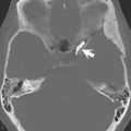
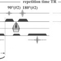
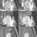
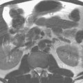
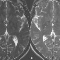
The images in Fig. 97.1 show the artifact (white arrows) from a nonferromagnetic aneurysm clip at 1.5 T. (A) Fast spin echo sequences have the least distortion due to the presence of multiple RF refocusing pulses. (B) is a diffusion-weighted image (see Case 63), which of all illustrated techniques, should show the greatest distortion. However, in this instance, parallel imaging has been employed with the result being substantially reduced artifact (but still greater than fast spin echo). (C) Gradient echo scans have very prominent artifact, due to the absence of a RF refocusing pulse. Three-dimensional time of flight MRA (see Case 43) uses a short TE and small voxels that decrease the area of the artifact, viewed on (D) the source image, when compared with (C) the 2D gradient echo scan. The distal right internal carotid artery (D
Stay updated, free articles. Join our Telegram channel

Full access? Get Clinical Tree


