Axillary Block

32 Axillary Block


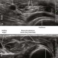
Three terminal branches of the brachial plexus (the median, radial, and ulnar nerves) lie close to the axillary artery in the axilla (Table 32-1). This makes the axilla a convenient place to block the brachial plexus (Table 32-2). Axillary block is traditionally performed by transarterial injection of local anesthetic around the axillary artery or by use of nerve stimulation to evoke motor responses. Transarterial block necessitates puncturing the axillary artery. Another weakness is failure to anesthetize the musculocutaneous nerve, which leaves the neurovascular bundle proximally underneath the pectoralis minor muscle at the level of the coracoid process.


Table 32-1 Characteristics of Terminal Branches of the Brachial Plexus in the Axilla


Nerve Characteristics
Axillary
Musculocutaneous
Radial
Median
Ulnar
Medial antebrachial cutaneous Lies between median nerve and ulnar nerve
Intercostobrachial and medial brachial cutaneous

Table 32-2 Clinical Considerations for Axillary Block with Ultrasound


Advantages Disadvantages


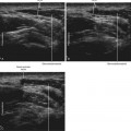
Ultrasound imaging improves axillary block of the brachial plexus. Almost all institutions have reported advantages to using ultrasound to guide this procedure.1,2 Ultrasound can be used to guide injections around the axillary artery. In addition, the musculocutaneous nerve can be directly imaged to complete the axillary block (see Chapter 33).


Suggested Technique


The transpectoral approach for proximal axillary block is performed with the needle tip just inside the chest, before the nerves of the brachial plexus diverge. With this lateral to medial approach, the needle enters through the pectoralis major muscle.


Axillary block is performed with the patient in supine position. The arm should be slightly hyperabducted to allow the needle placement to be as proximal as possible. Slightly more than 90 degrees of abduction is optimal for probe positioning. Because the pectoralis major inserts on the humerus, hyperabduction of the arm reduces the pectoral ridge by retracting the pectoralis major toward the midline. A Mayo stand can be used to support the arm in this position. The operator should stand at the head of the bed to view the ultrasound display across the patient’s arm.


The pectoral ridge separates the needle entry point and the transducer for this proximal axillary block. This can allow for coverless imaging because the needle entry site is remote from the transducer. The skin preparation is over the pectoralis major muscle. Tilting and rotating the angle of the transducer slightly into the chest torso allow for more proximal imaging. The axillary veins can be used as a manometer to measure the amount of probe compression. The correct amount of pressure for this procedure just coapts the walls of the veins.

Stay updated, free articles. Join our Telegram channel

Mar 5, 2016 | Posted by in ULTRASONOGRAPHY | Comments Off on Axillary Block

Full access? Get Clinical Tree

Get Clinical Tree app for offline access