The neuroradiological workup related to neuroophthalmology falls into the following categories based on anatomical lesion location and visual/cranial nerve pathways:
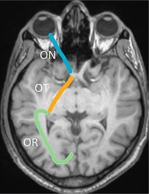
I. Visual/optic pathway – from the intracranial part of the optic nerves, optic chiasma, optic tracts, lateral geniculate body, optic radiations (including Meyer’s loop) and occipital (striate) cortex (Fig. 3.25.88)
II. Brainstem – involving the nuclear and internuclear substrates of the cranial nerves (III, IV and VI)
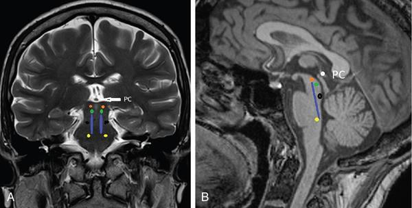
• Supplying the extraocular muscles (EOM) from cranial to caudal are (Fig. 3.25.89) – rostral interstitial nucleus – midbrain close to posterior commissure
• Medial longitudinal fasciculus (MLF) – from midbrain to pons
• Third nerve nucleus – upper midbrain – periaqueductal at superior collicular level
• Fourth nerve nucleus – lower midbrain at the level of superior cerebellar peduncles
• Sixth nerve nucleus – pons – under the facial colliculi in the floor of fourth ventricle
• Paramedian pontine reticular formation (PPRF)
III. Individual cranial nerves in basal subarachnoid space/cisterns
The three cranial nerves supplying the EOM have 3 segments as they course; cisternal – after exiting the brainstem, in the subarachnoid space.
Dural cave/interdural – along the walls or within the cavernous sinus (CS).
Intraorbital – intraconal space to supply the muscles.
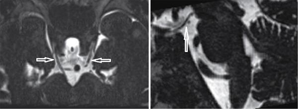
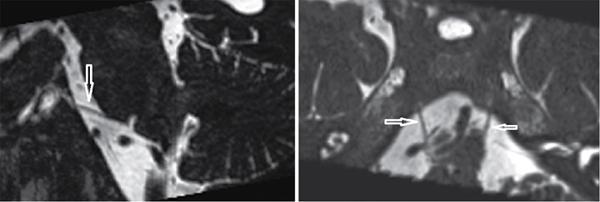
Oculomotor nerve – exits midbrain into the interpeduncular cistern, courses between the posterior cerebral artery (PCA) and superior cerebellar artery (SCA) and then courses close to the posterior communicating artery (PCom) and supraclinoid internal carotid artery (ICA) – to enter the cavernous sinus (Fig. 3.25.90).
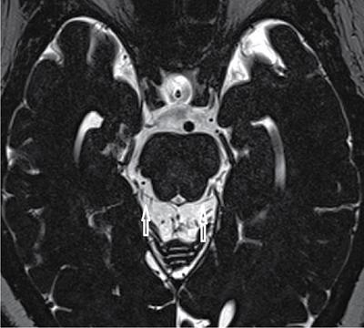
Trochlear nerve – exits on dorsal aspect of midbrain close to superior medullary velum into the quadrigeminal (tectal) plate cistern and courses around the midbrain in the ambient cistern – to enter the trochlear groove below the tentorium and reaches the cavernous sinus (Fig. 3.25.91).
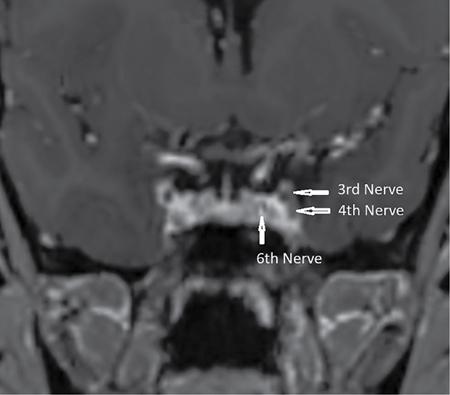
Abducens nerve – exits the lower anterior paramedian pons and ascends in prepontine cistern to enter the Dorello’s canal in the bony clivus to reach the cavernous sinus (Fig. 3.25.92).
IV. Cavernous sinus and orbital apex/SOF (Fig. 3.25.93)
• Third and fourth nerves along the lateral wall, sixth nerve within the CS (similar to the ICA)
• Third nerve divides into superior and inferior branches just before entering the SOF
• Enters the intraconal space within the annulus of Zinn
• Superior branch supplies the SR-LPS and inferior branch supplies MR, IR, IO
• Fourth nerve – enters the orbit outside the annulus of Zinn – supplies the superior oblique muscle
Heavily T2-weighted (CISS/SPACE/FIESTA/VISTA 3D) – nerves in the basal cisterns post-Gd (with fat-saturated FS) with or without MT (magnetization transfer)