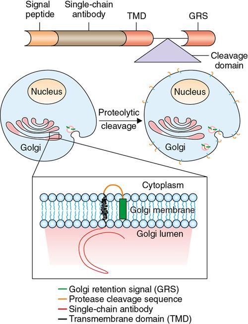
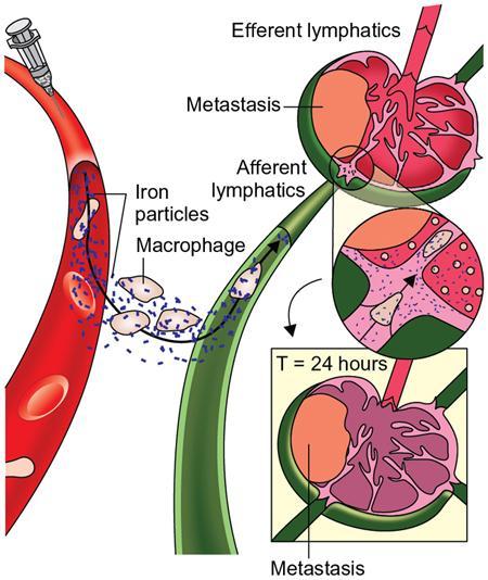
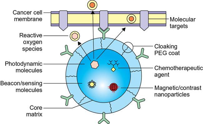
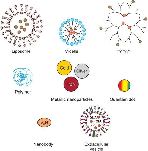
Sikandar Shaikh The blooming of a flower is, in my mind, not a miracle. It is something that we can understand on the basis of molecular biology these days. Francis Collins Molecular imaging is defined as the noninvasive imaging of cellular and subcellular levels. Molecular imaging is the fastest developing imaging modality at present with blend of applications from the existing imaging modalities, which are advanced, and this is leading to the concept of personalized medicine. The molecular imaging has become important in recent years due to the extensive research happening in the various clinical disciplines in every aspect. The advances in research in engineering, molecular biology, chemistry, immunology, medicine and genetics and combined with the multi- and interdisciplinary innovations of various modalities have resulted in this fascinating modality, which has diagnostic as well as therapeutic applications. The novel concepts of the molecular imaging have impacted for the earlier and better diagnosis. This is possible because of the noninvasive imaging modalities and newer protocols, which resulted in the earlier diagnosis, risk assessment and evaluation in relation to the monitoring to therapy. This had a significant impact on the high sensitivity and specificity and further resulted in better impact on overall management and also the various factors which will have an impact. Molecular imaging is the form of the functional imaging modalities such as positron emission tomography–computed tomography (PET-CT) which are newer one with many advancements, leading to earlier and better evaluation of the various disease pathologies which were not completely evaluated or not sensitive for evaluation by existing imaging modalities. The best examples are various neoplastic and newer applications such as infection and inflammation. This has answered many queries or doubts which were not cleared by existing modalities, such as size, extent, various organs involvement, various treatment options, monitoring response to treatment and most important side effects on the patient. Newer imaging modality such as PET-CT is commonly used worldwide for evaluation of neoplasms, infection and inflammation. Thus, PET, PET combined with CT and single-photon emission computed tomography (SPECT) are usually used. Technological advancements in magnetic resonance (MR), optical imaging, CT molecular imaging and ultrasonographic (US) imaging will be more widely used. CT is not a molecular imaging modality, but lot of research is being done with use of the X-ray attenuation and in future will be used as molecular imaging modality. Novel newer molecular imaging modalities are having great potential to significantly enhance the diagnostic and therapeutic approaches for cancer diagnosis and treatment. Molecular imaging techniques are having great potential to upgrade the management of cancer care by early detection and management of the various cancers. More specifically, oncologic molecular imaging is based on distinctive characterization of various molecular aspects of malignant cells. There are a lot of various biomolecules which has been developed, extensively studied and characterized and individual expression profiles have been defined for identification of the various types of cancer. Molecular imaging probes which are used to target and highlight these specific characteristics can be evaluated directly or indirectly by the various cells in vicinity such as T cells, macrophages, dendritic cells, fibroblasts or endothelial cells (Fig. 1.19.1). Recently molecular imaging modalities, which are used as whole-body imaging modalities for cancer diagnosis and staging, are US, CT and MR with newer concepts by expanding morphological imaging to a cellular and functional imaging. The precise evaluation of cellular properties of microenvironments is characteristic for the malignancy. This will be enabling earlier detection and assessment of more personalized management approach. The most widely used molecular imaging modality is PET with 18F-fluorodeoxyglucose (18F-FDG), which diagnoses the metabolic discrepancies between malignant and normal cells, making PET the important and widely used molecular imaging modality. The technical drawbacks are related with high cost, use of ionizing radiation and low spatial resolution which restricts its potential. Molecular imaging with higher-resolution modalities such as magnetic resonance imaging (MRI) is being more widely used, and a lot of advancements will also be coming. Molecular imaging probes are also being used for earlier diagnosis especially for the solid cancers, thus facilitating the better management options. Molecular imaging techniques have significant potential to enhance cancer diagnosis and staging on multiple levels, especially in relation to tumor detection and characterization. US, CT and MRI shows continuous advancement in the techniques the tumor detection is being more precise and focused. Thus, the basis of the molecular imaging applications is to make carcinogenesis visible at the earliest as alterations on the cellular level are targeted and detected very soon. The pathogenesis of the tumors and inflammation is based on the fluid in the extracellular space, which is causing the endothelial disruption and ultimately resulting in the end-stage organ damage. The response to the injury in these pathologies results in the accumulation of leukocytes macrophages, monocytes, lymphocytes and giant cells in the area of the pathology. This mechanism will result in the activation of the various secretions of the inflammations such as cytokines and chemokines. Nowadays, PET-MR is being considered as the modality of the choice especially in the grey areas where PET-CT is of less relevance. PET-MR outnumbers in few of these pathologies such as collagen disorders, various infections in the body, fever of unknown origin, vasculitis and many conditions where soft-tissue evaluation is more focused. Still many conditions where subtle or borderline imaging findings are not being evaluated by these newer imaging modalities. These include mostly the pediatric diseases, vascular pathologies, disseminated infections in the body, etc. The hallmark of any infective and inflammatory pathologies is the role of macrophages and monocytes especially in the Inflammations. Recent advances have shown that the imaging of these cells can be done by using the more specific small and ultrasmall paramagnetic iron oxide nanoparticles (SPIO and USPIO). The principle here is phagocytosis principle and includes the process of surface coating and opsonization. The amount of uptake is again based on the size, surface coating and specific ligands pathways for particular molecule or biomarker. These also can be evaluated by using more specific PET and SPECT ligands and also fluorescence imaging. The immune system is based on the various factors impacting the B lymphocytes especially in relation to the hormonal immunity by the antibody response in response to the various antigens. These B cells are also labelled as cell markers for specific cells. These B cell functioning has resulted in diagnosing the various disease patterns especially autoimmune diseases. One of the biomarkers is the rituximab, which is the radiolabeled marker for identifying the immune response to the pathologies such as rheumatoid arthritis, psoriatic arthritis and sarcoidosis, thus predicting the wider role in the identification of the radioimmunotherapies in various conditions. T cell lymphocytes imaging is important for the evaluation of the cell-mediated immune response especially in identifying the various inflammation and autoimmune disorders. With this probability, many new radiotracers are being tried for various indications and pathologies in relation to aforementioned conditions. Cytokines which are radiolabelled are one of the important ones and more commonly used. The various applications related with the cancer imaging are attributed to the wide range of the modalities available. As mentioned, it does have important role in the evaluation of various aspects of the cancer management right from the diagnostic options to various therapeutic options. As already mentioned, and described the role of the various imaging modalities, every modality has its relevance related to the applications in which they are more precise, to evaluate leading to the concept of the personalized medicine. Nanomaterials are also being the one of the common imaging modalities for the imaging of the cancer. The use of the targeted molecular imaging probes is being tried for CT. The use of the bismuth sulphide nanoparticles can provide better sensitivity than iodine-based probes due to their higher atomic numbers, and at the same time, some limitations of iodine-based contrast agents can be overcome. Nanoparticle-based molecular imaging probes for CT are showing excellent results for both the imaging of the cancer and infection and inflammation in future. Inflammation of the central nervous system is seen in many conditions such as autoimmune syndrome, various age-related degenerative conditions, various neoplastic conditions and psychiatric disorders. The main concept of the evaluation is the use of various receptors which are specific targets to various proteins, different membranes and macrophages of various nature, and these are the important targets for evaluation of the imaging in the neuroinflammation. One of the excellent examples is the use of the iron oxide magnetic nanoparticles for the infarct evaluation in various stages. Magnetic nanoparticles use is seen as more specific and focussed than the use of gadolinium in conditions such as multiple sclerosis. Vascular pathologies are important conditions quite common in this older age group, and evaluation of the atherosclerosis where the plaque development risk is more can be evaluated with the macrophage activity. This can also identify the factors where more risk of the plaque is there and also the chances of the rupture can also be identified. One of the important imaging modalities is the 18F-FDG PET-CT, which is evolving as an important modality for imaging the various vascular plaques especially vascular and associated macrophage activity in the region of the interest. Some of the biomarkers used are TSPO expression and radiolabelled DOTATATE. In atherosclerosis, the use of the SPIO nanoparticles is also being well established so that early evaluation can be more beneficial for the outcome of the morbidities. The evaluation of the various infections and inflammations in the lung parenchyma can be done with various biomarkers and is being well established, and the same concept applies to the evaluation of the abdominal pathologies. The role of the 18F-FDG PET-CT is now well established in various conditions such as radiation pneumonitis, congenital conditions such as cystic fibrosis, and bronchial asthma. There is also established role in the evaluation of the myocardial ischaemia and infarction by using the USPIOs, and this will also be used in future for the evaluation of the inflammatory cells. Still lot needs to be done to consolidate this. The role of the 18F-FDG PET-CT is now clearly defined as an important modality for the evaluation of the various inflammatory arthritis in different joints. Further the role of various imaging modalities with more specific diagnostic criteria is also being done by using the 18F-FDG PET and USPIO in various conditions of the various collagen disorders. Fluorophores as an optical imaging agent form the basis of use of an optical signal, resulting due to the excitation light source of specific wavelength; this is the basic principle of the optical imaging. Indocyanine green is one of the agents added to the target molecule for imaging. The emitted signal is captured by device camera. The biggest advantage of optical imaging is to evaluate fast imaging with high-resolution images. The advantage is more flexible and can be used more often for imaging. The biggest disadvantage is that the signal is extremely poor with depth of 1 centimeter and thus can be used only for imaging of the superficial tissues only. Optoacoustic and photoacoustic imaging are the newer techniques which are having more penetration depth compared with the routine optical imaging and thus can be used for more wider applications having more depth requirements. Thermal expansion of a tissue forms the basis of the optoacoustic imaging, and this can be done by using intrinsic fluorophores or any another agent with similar properties and cause shorter pulses. This thermal expansion is caused by ultrasonic fissures, which are generated due to the more specific waves by using specific agents and one of these are fluorophores. This imaging has incredibly good penetration depth of 3–5 cm approximately. Due to this principle, the optical and optoacoustic imaging are being more widely used compared with the existing modalities such as PET, SPECT, MR and ultrasound. PET-MR being the hybrid imaging modality has more sensitivity to characterize various tissues and can do better physiological localization within the soft tissues. The advantages of the PET-MR are more in the various soft-tissue pathologies such as spondylodiscitis, various infections and inflammations in different parts of the body. The use of the molecular ultrasound by use of the various molecular agents will be having significant impact on the diagnostic and therapeutic ultrasound in the future. The use of the thermal energy forms the basis of the therapeutic ultrasound for treatment of various pathologies along with the drugs formulations. Few of the commonly used molecular ultrasound agents are microbubbles, targeted agents and carriers or enhancers of drug related with drug release. These have resulted in increasing the efficacy of the drug target and drug release easily, which was a big challenge. Ultrasound molecular imaging has become well-established modality with the great advantage that it is noninvasive and no radiation. Due to this, it has become the modality of choice for many clinical applications. The advancements in the ultrasound contrast agent’s development have revolutionized the molecular imaging concept. The various applications of the ultrasound are based on the changes due to various contrast agents and markers on various tissues such as vascular endothelium. There is a significant role in the evaluation of the angiogenesis, inflammation and thrombus formation in the vessel wall. This is possible due to newer ultrasound contrast developments, ultrasound technology that has resulted in the significant improvement in the sensitivity of various applications. The biggest advantage of ultrasound is the easy availability and very low cost compared with other modalities where it can be repeatedly used in paediatric age groups, various malignancy settings, etc. MRI as an imaging modality has an excellent soft-tissue resolution, which gives more precise information about the tissue architecture. The biggest advantage is no radiation. MRI-based molecular imaging targeting agents are not as sensitive as that of agents which are used in PET and SPECT due to their little, bigger size. Magnetic nanoparticles have the ability to disturb the relaxation of nearby protons which can be detected by MRI (Fig. 1.19.2). Nanoparticles can be used to detect vascular leak, and this forms the basis for evaluation of the inflammation. Nanoparticles are phagocytosed by macrophages, and with this basis, IO-containing formulations will cause a darkening of the reticuloendothelial system in the liver and spleen on T2-weighted images. This has been used to detect various inflammations in patients with renal transplant rejection, atherosclerosis and multiple sclerosis. The precise and noninvasive evaluation of endothelial inflammation with MRI and VCAM-1 targeted MPIO can help in diagnosis of various conditions such as atherosclerosis, renal and cerebral parenchyma evaluation. The other conditions which can be detected are focal brain ischaemia, blood–brain barrier breakdown and brain inflammation. Thus, MRI has become an important imaging modality for the evaluation of various inflammations in the different organs by using various MRI-based agents. This has resulted in excellent anatomic delineation and also the functional aspects by using various molecular agents. The nanoparticle contrast agents are the mainstay in the imaging and had resulted in the nanoimaging. Thus, nanoimaging is being used widely now for various pathologies evaluation of various pathologies. This is possible due to the significant technical and probe design advancements. The probes which are designed are for having multiple functions having capacity to detect better targets, more compatibility in the biological tissues so that they are safe and also have maximum diagnostic and therapeutic properties (Fig. 1.19.3). For this, we should have an ideal nanoparticle, and they should be formulated in such a way that it should be very easy to use, have better compatibility in tissues, better efficiency, increased specificity and sensitivity, and be able to detect disease entity in more precise manner. One of the important applications is in cancer imaging for diagnosis and therapeutics. Various nanoparticles are being used for different applications. Carbon nanotubes are one of the commonly used nanoparticles; they are made up of the sheets of carbon atoms which are rolled in the form of the cylindrical shape of one atom thickness. DNA mutations and proteins can be easily identified by this technique. The thermal principle of generating the heat by exposure to the radiofrequency fields forms the basis of the imaging. Dendrimers are also the important imaging particles which are structured as branched molecules. These dendrimers are used along with the various other agents such as folic acid, and gold nanoparticles are used for the evaluation of cancer cells. The sustained controlled release system is also important hallmark for better and uniform drug delivery. The therapeutic options also are based on the photodynamic–thermal therapy principle. Many nanoagents used are gadolinium, various radionuclides with specific applications, chemotherapeutic agents, photodynamic therapeutic agents and reactive oxygen; these are few for various therapeutic options (Fig. 1.19.4).
1.19: Basics and advances in molecular imaging
Basics in molecular imaging
Introduction
Pathophysiology
Tumours
Infection and inflammation
Imaging of macrophages and monocytes
Imaging of lymphocytes
Imaging of the cancer molecular imaging
Inflammation imaging
Inflammation of central nervous system
Imaging of vascular pathologies
Imaging of thorax, abdominal organs and joints
Optical imaging
Ultrasound molecular imaging
Magnetic resonance molecular imaging
Nanoimaging
Stay updated, free articles. Join our Telegram channel

Full access? Get Clinical Tree


