Basics of CT Scanning and Issues Relevant to Integrated Imaging
Willi A. Kalender
X-ray computed tomography (CT) has evolved to be one of the most important imaging modalities due to the introduction of spiral CT and of multi-row detector technology. Multi-slice spiral CT (MSCT) allows coverage of complete organs in typically 5 to 10 seconds or “whole-body” scans in less than 30 seconds with isotropic spatial resolution of typically 0.5 mm or better. This chapter focuses on technical developments, scanner performance, image quality, and the expanded range of clinical applications. Dose issues are dealt with briefly, in particular, the options for dose reduction offered by modern CT. MSCT constitutes a perfect match with molecular imaging using PET and SPECT. Given the current high performance levels, the latest advances in CT technology, including cardiac CT, can and should be utilized without any restriction in PET-CT and SPECT-CT scanners.
Introduction
X-ray computed tomography (CT) has gone through a number of development cycles since its introduction in the early 1970s. Although the goals and the future of CT developments were rated differently at different points in time, in particular when compared with competing modalities, such as magnetic resonance imaging (MRI), that emerged in parallel, a general trend has been observed throughout these years: it has been a primary aim to speed up measurements, both for the scanning of single slices and for the scanning of volumes. The introduction of spiral CT scanning in 1989 was a decisive step in this direction and has led to a renaissance of CT: CT is largely regarded as equivalent to spiral CT today. Since the end of the 1990s, rotation times of 0.5 seconds and below and multi-row detectors have become available. Today they allow measuring up to 64 slices simultaneously. In combination with the short rotation times, this provides for unprecedented volume scan times: single-organ examinations take less that 10 seconds, and whole-body examinations can be completed in less that 30 seconds. CT is a universal imaging modality covering all organs and body regions; even cardiac imaging, including CT coronary angiography, is performed routinely in spiral mode today. As an illustration of the performance levels, Figure 6.1 shows typical results achieved with 64-slice scanners. The introduction of “dual source CT” (DSCT) systems—scanners with two x-ray tubes and two detectors intended to improve cardiac CT and provide dual-energy CT—is the latest of many recent innovations.
This chapter offers a short review of the principles and technology involved in modern CT, an assessment of its performance in clinical imaging as a balance of image quality and dose, a brief overview of special applications, and a statement regarding the suitability of modern CT technology for combination scanning with positron emission tomography (PET) and single-photon emission computed tomography (SPECT).
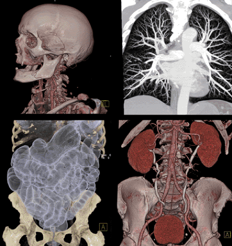 Figure 6.1 Multi-slice spiral CT scans cover all body regions in a matter of seconds. Even “whole-body” scans are obtained in less than 30 seconds. The examples shown were obtained on a 64-slice scanner with 0.33-second rotation time and 32 × 0.6 mm collimation and double sampling. (Courtesy of Werner Bautz, University of Erlangen, Erlangen, Germany.) |
Spiral CT Principles
Traditionally, CT images were acquired by sequential scanning of single slices; 3-D volumes were built up sequentially from such 2-D scans. In spiral scanning, the patient is transported continuously through the field of measurement while the x-ray tube and the detector rotate continuously (1,2). Relative to the patient, the x-ray focus travels on a spiral path (Fig. 6.2A). The technological basis, continuous rotation of the measurement system, became available in the late 1980s and is now generally used in all types of scanners, in high-performance as well as in low-cost scanners, and also in combination scanners used for PET-CT and SPECT-CT. The spiral scan mode provides two major principal advantages: (a) decisively faster volume coverage than sequential slice-by-slice scanning would ever allow and (b) increased 3-D resolution, both for low-contrast and for spatial resolution, due to the continuous data sampling along the z-axis (1).
Image reconstruction is the same in principle for sequential CT and for spiral CT. However, in spiral CT the data sets representing a single slice have to be generated from the spiral volume data set in an additional preprocessing step, the so-called z-interpolation. This step is not
computationally expensive but it provides a decisive advantage: images can be reconstructed for any arbitrary position. Both the position of the image and the effective slice width can be chosen retrospectively. Reconstructing overlapping images is the basis for improved low-contrast and high-contrast resolution. Details on aspects of image reconstruction in spiral CT are given elsewhere (1) but are not necessary for understanding this chapter.
computationally expensive but it provides a decisive advantage: images can be reconstructed for any arbitrary position. Both the position of the image and the effective slice width can be chosen retrospectively. Reconstructing overlapping images is the basis for improved low-contrast and high-contrast resolution. Details on aspects of image reconstruction in spiral CT are given elsewhere (1) but are not necessary for understanding this chapter.
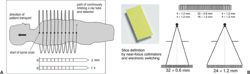 Figure 6.2 Multi-slice spiral scanning using array detector technology is the method of choice in modern CT. A: The spiral scan principle (2). B: An example of a multi-row detector with 40 rows for simultaneously acquiring 32 slices or 64 slices when double z-sampling is available. (Reprinted with permission from Kalender WA. Computed Tomography. Erlangen: Publicis Corporate Publishing; 2005. ) |
Technology
Many technical components of a modern CT scanner with subsecond rotation times represent cutting-edge technology and are constantly upgraded. Detector technology was the focus of interest for many years, as the detector was viewed as the most important component, and new concepts for its design have made multi-slice scanning possible and have advanced CT to its present level. But other components are gaining in importance too.
The importance of the mechanical setup increases with shorter and shorter rotation times. For rotation times of 0.3 seconds, centrifugal forces of about 30 g result, which are far beyond what humans could tolerate. For a mechanical system, it would be possible to go beyond the typical rotation times of 0.3 to 0.4 seconds currently offered by the top-performance systems. However, not only would adapting the components involved (e.g., the rotating-anode x-ray tube) involve added expense, there is the problem of available x-ray power, which is the main limiting factor. The numbers of photons necessary to generate an image with acceptable quality is fixed and has to be provided in shorter and shorter time intervals when the rotation speed is increased. This limitation, which was an essential reason for the failure of electron-beam CT, has to be kept in mind. It provided the motivation to think about multi-source systems again, which are addressed below.
The main improvements in x-ray tube technology for CT over the years consisted of increasing the mass and the diameter of the rotating anode to allow higher and continued power levels. Today, the most powerful CT systems offer 80- to 100-kW x-ray sources. An important technological innovation has been introduced just recently, the so-called rotating vacuum vessel technology (1,3). In this technology, the complete vacuum vessel, held by dual bearings, rotates, and the anode is an integral part of the vessel (Fig. 6.3A). This allows for direct cooling of the anode’s back surface and alleviates the need for a large and massive anode. This new technology offers an additional advantage: since the electron beam has to be steered electromagnetically from the centrally placed cathode to the anode, the focus position can be controlled and switched precisely. This feature is utilized by generating a so-called z-flying focal spot, which allows for double-sampling in the z-direction and has beneficial effects on resolution and artifact behavior (1,4). A tube of this design, the Siemens Straton tube, is shown disassembled in Figure 6.3B to illustrate the reduced mass and dimensions. The anode has a diameter of only 12 cm but allows 80-kW power levels. It has been in successful use in clinical CT for more than 2 years. Such designs will likely become preferred for CT x-ray sources in the future.
Related posts:
 11C-Based PET- Radiopharmaceuticals of Clinical Value
 Brain Tumors: Astrocytoma, Oligodendroglioma, and Glioblastoma Images with PET and SPECT
 Sentinel Node Imaging in Head and Neck Tumors
 PET-CT and SPECT-CT of Bone Metastases
 SPECT and PET in Benign Thyroid and Parathyroid Disease
Stay updated, free articles. Join our Telegram channel

Full access? Get Clinical Tree


