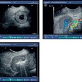
1 Basics of Radial Endoscopic Ultrasound Endoscopic ultrasound today demands the highest level of expertise from physicians working in endoscopy. The method requires a combination of familiarity with endoscopic techniques and experience in ultrasound. When it is properly performed and critically assessed, it makes extensive diagnostic and therapeutic applications possible. However, newcomers to the field often find themselves facing a multitude of problems, even when they have sufficient knowledge of endoscopy and sonography. The consequent frustration can result in this valuable method being abandoned. This chapter therefore aims to simplify the examination technique so that it can be carried out successfully after a little practice. Firstly, we can introduce the “ten golden rules of endoscopic ultrasound,” established by the endoscopic ultrasound specialist Dr. Uwe Will and confirmed in practice by other experienced endosonographers. These rules should be remembered when difficulties arise during an examination (in both radial and longitudinal ultrasound), and they make it possible to regain orientation: The patient should be made aware of the risks and effectiveness of the procedure beforehand. Endoscopic ultrasound is not suitable as a screening method and should only be used to answer specific questions. In principle, the procedure used in the examination depends on the question being posed. For the newcomer, the easiest phenomena to examine and diagnose are protuberances in the digestive tract and surface tumors. The instrument is positioned directly in front of the relevant area, and the examination takes place under continuous suction. However, although this process is easy to carry out in the duodenum, antrum, and esophagus, the presence of air and the many folds in the gastric body and fundus regions make it more difficult there. The most difficult areas to examine are the angular notch and the fundus region. Small protuberances in the gastrointestinal tract are often difficult to image with the balloon method alone. The examiner should be aware that mucosal masses may be “flattened out” with the balloon and therefore remain hidden. These should preferably be examined using the miniprobe method, which makes it possible to locate lesions more precisely. The most difficult endoscopic ultrasound procedure is examining extraintestinal organs and structures. Success here depends on excellent knowledge of ultrasound anatomy, so that pathological structures can be detected by recognizing markers. As a rule, the operator should make sure that the radial endoscopic ultrasound instruments are standardized for the “CT view”—i.e., viewing the patient from underneath. This can best be seen in the mediastinum. It is essential to remember that when the descending aorta and the spine are at the bottom edge of the picture, the image will appear mirrored: the right main bronchus appears on the left side of the image, the left main bronchus on the right. To maintain orientation during the imaging process, it is necessary to identify a series of standard ultrasound cross-sections in the gastrointestinal tract. This should be performed during every examination. Each cross-section and the best technique for locating it are described below. To produce images of extraintestinal structures, one should start by positioning the scope in the descending duodenum by sight in the same way as in endoscopic retrograde cholangiopancreatography (ERCP), and then straighten it slowly. Using the water-filled balloon is recommended in order to improve the contact (preferred by one of the present authors, M.H.), but as mentioned before, some practitioners do not consider this necessary (preferred by C.F.D.). The left hand is used to maintain continuous suction and also to angle the tip of the scope to optimize contact with the intestinal wall. Sideways movements are made by turning the instrument with the right hand, or by turning the whole instrument at the shaft. The small wheel is not usually used for positioning. As the scope is pulled back slowly from this relatively blind position, two anatomic markers become visible: the inferior vena cava and aorta on the left side of the screen and the superior mesenteric vein on the right side. These two markers form a “V” shape, which surrounds parts of the pancreas. This is the first recognizable image to appear while the scope is being withdrawn from the descending duodenum; the view of the pancreas opens up—the “golden V”—when this position has been reached (Fig. 1.1).
 Cross-Section of the Descending Duodenum
 Notes
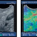
Notes Cross-Section of the Papilla of Vater
Related posts:


Stay updated, free articles. Join our Telegram channel

Full access? Get Clinical Tree


