A BI-RADS (Breast Imaging Reporting and Data System) 3, or probably benign, assessment is given in approximately 7% to 12% of breast magnetic resonance (MR) images. However, the imaging features of probably benign lesions on MR imaging have not been well defined. As with mammography and ultrasonography, a BI-RADS 3 assessment should be used only when there is a less than 2% likelihood of malignancy. The use of BI-RADS 3 for classically benign findings should be avoided. Certain masses, foci, and areas of nonmass enhancement may be categorized as probably benign on baseline MR imaging.
Key points
- •
A BI-RADS (Breast Imaging Reporting and Data System) 3, or probably benign, assessment is given in approximately 7% to 12% of breast magnetic resonance (MR) images.
- •
As with mammography and ultrasonography, a BI-RADS 3 assessment should be used only when there is a less than 2% likelihood of malignancy.
- •
The use of BI-RADS 3 for classically benign findings (ie, chronic cysts, classic fat necrosis, intramammary lymph nodes, and multiple bilateral similar benign-appearing masses) should be avoided.
- •
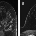
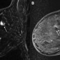
The most appropriate uses of a probably benign assessment on MR imaging include benign-appearing foci that are unique and distinct from the background enhancement or masses typical of incidental benign fibroadenomas.
- •
Short-term follow-up may also be appropriate for areas of benign-appearing focal nonmass enhancement that probably represent variations of the normal background enhancement.
The use of the probably benign category for lesions detected on breast magnetic resonance (MR) imaging is not standardized. Compared with the well-established criteria for assessing a mammographic or sonographic finding as probably benign, the data describing the appropriate use of the probably benign category on MR imaging are limited. Therefore, the approach to categorizing findings on breast MR imaging as probably benign has been borrowed primarily from principles used in the evaluation of mammographic lesions. This approach involves the analysis of lesion morphology, distribution, and symmetry. However, MR imaging assessment also incorporates additional information such as water content (T2 signal intensity) and kinetic analysis.
The American College of Radiology (ACR) Breast Imaging Reporting and Data System (BI-RADS) is used to classify breast imaging studies to indicate the level of suspicion for malignancy for any given study. BI-RADS categories, originally established for mammography, are classified as normal (BI- RADS 1), benign (BI-RADS 2), probably benign (BI-RADS 3), suspicious (BI-RADS 4), highly suggestive (BI-RADS 5), or known malignancy (BI-RADS 6). Nonpalpable, probably benign BI-RADS 3 lesions, which are reported in 1% to 11% of mammograms, have imaging features that suggest a less than 2% chance of malignancy. The imaging criteria of BI-RADS 3 mammographic lesions are largely based on 2 studies that collectively include more than 80,000 mammograms. Examples of mammographic probably benign lesions include an incidental low-density circumscribed mass or a cluster of punctate calcifications on a baseline mammogram. In these studies, a 6-month follow-up mammogram detected interval progression of those few BI-RADS 3 lesions that were malignant but still diagnosed these cancers early enough to maintain a favorable prognosis. Close follow-up of BI-RADS 3 lesions reduced the false-negative rate of biopsy, decreasing health care costs.
Because of the increasing use of MR to evaluate the breast, in 2003 the ACR expanded BI-RADS to include a lexicon describing findings on breast MR imaging. Although not required by the Mammography Quality Standards Act, the ACR recommends using BI-RADS final assessment codes for breast MR imaging examinations. Under the BI-RADS for MR imaging, a BI-RADS 3 assessment is defined as the following:
A finding placed in this category is highly unlikely for malignancy and should have a very high probability of being benign. It is not expected to change over the follow-up interval, but the radiologist would prefer to establish its stability. Data are becoming available that shed light on the efficacy of short-interval follow-up. At the present time, most approaches are intuitive.
In this article, the published literature on BI-RADS 3 lesions on MR imaging is reviewed, and the imaging features of lesions that may appropriately be categorized as probably benign are discussed.
Frequency and cancer yield
Few studies are available in the literature regarding the frequency of use of BI-RADS 3 assessments and the cancer yield on follow-up imaging. With the exception of 1 study, short-term follow-up was recommended in approximately 6% to 12% of examinations ( Table 1 ). One of the earliest studies addressing the imaging features and outcomes of BI-RADS-3 lesions was a retrospective study by Liberman and colleagues published in 2003. In this study, 24% (89/367) of baseline studies in high-risk women received a BI-RADS 3 assessment. This high frequency of BI-RADS 3 assessment may reflect the learning curve of interpreting radiologists and the benefit of having comparison studies. Warner and colleagues reported a decrease in the frequency of BI-RADS 3 recommendations from 8% on baseline studies to 3% on subsequent rounds of screening.
| Reference | Type | Study Population | Probably Benign Examinations (Number [%]) | Probably Benign Patients (Number [%]) | Cancer Yield (Number [%]) |
|---|---|---|---|---|---|
| Kuhl et al, 2000 | Prospective | High risk | 45/363 (12.4) | 44/192 (22.9) | 1/44 (2.3) |
| Liberman et al, 2003 | Retrospective | High risk | 89/367 (24.2) | 89/367 (24.2) | 9/89 (10.1) |
| Kriege et al, 2004 | Prospective | High risk | 275/4169 (6.6) | NR/1909 | 3/275 (1.1) |
| Hartman et al, 2004 | Prospective | High risk | 19/75 (25) | 14/41 (34.1) | 0/14 (0.0) |
| Sadowski & Kelcz, 2005 | Retrospective | BI-RADS 0 mammogram | NR | 79/473 (16.7) | 4/68 (6) |
| Kuhl et al, 2005 | Prospective | High risk | 167/1452 (11.5) | NR/529 | NR |
| Eby et al, 2009 | Retrospective | High risk, extent of disease | 260/2569 (10.1) | 236/1735 (13.6) | 2/236 (0.9) |
| Weinstein et al, 2010 | Prospective | Known contralateral cancer | 106/969 (10.9) | 106/969 (10.9) | 1/106 (0.9) |
One of the largest studies evaluating probably benign lesions is by Eby and colleagues. Of the 362 lesions included, 168 (46%) were foci, 132 (36%) areas of nonmass enhancement (NME), and 62 (17%) masses. In the prospective multicenter American College of Radiology Imaging Network (ACRIN) 6667 trial, a total of 145 probably benign lesions were followed, including 47 (32%) foci, 59 areas of NME (41%), and 37 (26%) masses.
The cancer yield of probably benign lesions has ranged between 0% and 10%. Liberman and colleagues found that 10% (9/89) of lesions initially characterized as probably benign subsequently were found to represent malignancy, which is substantially greater than the less than 2% frequency of malignancy that is considered acceptable for probably benign lesions on mammography. One reason may be that this study helped to identify certain types of lesions, such as NME in a ductal distribution, which warrant biopsy and should not be categorized as BI-RADS 3 lesions.
Since this initial study, additional retrospective and prospective studies have evaluated the frequency and outcomes of probably benign lesions on breast MR imaging. Both the frequency (7%–25% of examinations) and cancer yield (0%–5%) in these studies vary widely, which may partly be caused by differences in the inclusion criteria of the study population as well as differences in the criteria for what constitutes a probably benign lesion. In 1 prospective multi-institutional trial, 11% of women received a BI-RADS 3 recommendation and the frequency of malignancy in probably benign lesion was 0.9%.
Frequency and cancer yield
Few studies are available in the literature regarding the frequency of use of BI-RADS 3 assessments and the cancer yield on follow-up imaging. With the exception of 1 study, short-term follow-up was recommended in approximately 6% to 12% of examinations ( Table 1 ). One of the earliest studies addressing the imaging features and outcomes of BI-RADS-3 lesions was a retrospective study by Liberman and colleagues published in 2003. In this study, 24% (89/367) of baseline studies in high-risk women received a BI-RADS 3 assessment. This high frequency of BI-RADS 3 assessment may reflect the learning curve of interpreting radiologists and the benefit of having comparison studies. Warner and colleagues reported a decrease in the frequency of BI-RADS 3 recommendations from 8% on baseline studies to 3% on subsequent rounds of screening.
| Reference | Type | Study Population | Probably Benign Examinations (Number [%]) | Probably Benign Patients (Number [%]) | Cancer Yield (Number [%]) |
|---|---|---|---|---|---|
| Kuhl et al, 2000 | Prospective | High risk | 45/363 (12.4) | 44/192 (22.9) | 1/44 (2.3) |
| Liberman et al, 2003 | Retrospective | High risk | 89/367 (24.2) | 89/367 (24.2) | 9/89 (10.1) |
| Kriege et al, 2004 | Prospective | High risk | 275/4169 (6.6) | NR/1909 | 3/275 (1.1) |
| Hartman et al, 2004 | Prospective | High risk | 19/75 (25) | 14/41 (34.1) | 0/14 (0.0) |
| Sadowski & Kelcz, 2005 | Retrospective | BI-RADS 0 mammogram | NR | 79/473 (16.7) | 4/68 (6) |
| Kuhl et al, 2005 | Prospective | High risk | 167/1452 (11.5) | NR/529 | NR |
| Eby et al, 2009 | Retrospective | High risk, extent of disease | 260/2569 (10.1) | 236/1735 (13.6) | 2/236 (0.9) |
| Weinstein et al, 2010 | Prospective | Known contralateral cancer | 106/969 (10.9) | 106/969 (10.9) | 1/106 (0.9) |
Related posts:
 Breast Magnetic Resonance Imaging Technique at 1.5 T and 3 T
 Updates and Revisions to the BI-RADS Magnetic Resonance Imaging Lexicon
Screening MR Imaging Versus Screening Ultrasound
Updates and Revisions to the BI-RADS Magnetic Resonance Imaging Lexicon
Screening MR Imaging Versus Screening Ultrasound
 Evaluation of Breast Silicone Implants
 Magnetic Resonance Imaging–Guided Biopsy of the Breast
 Radiologic-pathologic Correlation at Breast MR Imaging
Stay updated, free articles. Join our Telegram channel

Full access? Get Clinical Tree


