12 Bladder, Prostate, and Uterus
Up to now we have deliberately focused our attention on the organs of the upper abdomen that are classic subjects for examination by ultrasound. However, every ultrasound examination of the abdomen should also include a look at the organs that fall within the domain of urology and gynecology. It is important, therefore, to give a brief account of the bladder, prostate, and uterus.
 Organ Boundaries and Relations
Bladder and prostate
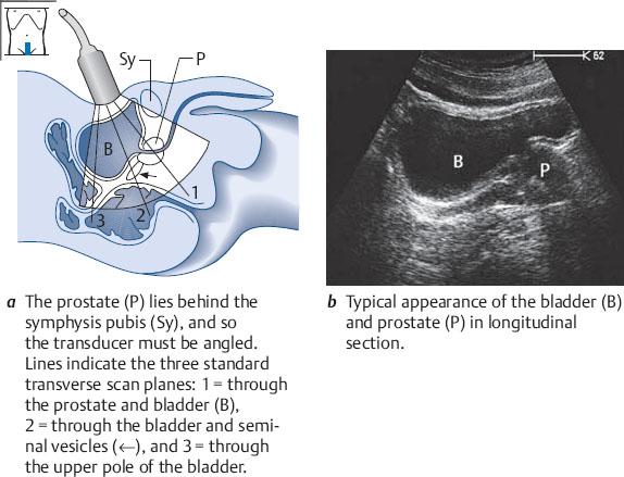
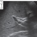
Demonstrating the bladder and prostate in longitudinal section
Place the transducer longitudinally in the midline just above the symphysis pubis, preferably with a full bladder. Angle the scan slightly downward. Identify the echo-free lumen of the bladder and, behind it, the prostate (Fig. 12.1). The prostate is hypoechoic, homogeneous, and bounded by a capsule. It normally measures up to 35mm in its craniocaudal dimension.
Demonstrating the bladder and prostate in transverse section
The transverse diameter of the prostate is 3–4 cm, its anteroposterior diameter is 2–3 cm, and its craniocaudal diameter is up to 3.5 cm.
The normal prostate is uniformly hypoechoic.
Scan transversely above the symphysis pubis and angle the probe sharply downward. Identify the bladder and prostate (Fig. 12.2a
Related posts:
Stay updated, free articles. Join our Telegram channel

Full access? Get Clinical Tree


