Usually from left side of distal thoracic esophagus
• Chest film
Left side pleural effusion or hydropneumothorax
Radiolucent streaks of gas along aorta or in neck
• Esophagography with nonionic, water-soluble contrast agent
Shows extravasation of ingested or injected (through nasogastric tube) contrast medium
From left side of esophagus, just above gastroesophageal (GE) junction
If initial study with water-soluble contrast medium fails to show leak, examination must be repeated immediately with barium to detect subtle leaks
• CT
Extraluminal gas &/or oral contrast medium in lower mediastinum &/or upper abdomen
TOP DIFFERENTIAL DIAGNOSES
• Mallory-Weiss syndrome
• Pulsion diverticulum (epiphrenic)
• Iatrogenic (postinstrumentation) injury
CLINICAL ISSUES
• Accounts for 15% of total esophageal perforation cases
• Prognosis for large perforation
After 24 hours without treatment: Mortality = 70%
After immediate surgical drainage: Good
• Treatment
Drains in esophagus, mediastinum, pleural space, &/or abdomen
(Left) Graphic shows a vertically oriented laceration of the distal esophagus, just above the hiatus and gastroesophageal (GE) junction.
(Right) Film from an esophagram following injection of a water-soluble contrast medium through a nasogastric tube demonstrates a leak of contrast medium from a tear in the left anterior wall of the distal esophagus , a classic appearance for Boerhaave syndrome.
(Left) Axial CECT in a middle-aged man with severe chest pain after repeated retching shows extraluminal gas and contrast material surrounding the esophagus in the lower mediastinum and upper abdomen.
(Right) Film from a fluoroscopic exam in the same patient during injection of water-soluble contrast through a nasogastric tube shows extraluminal contrast in the mediastinum and upper abdomen . The site of the tear is the left anterior wall of the distal esophagus.
TERMINOLOGY
Definitions
• Spontaneous distal esophageal perforation following vomiting or other violent straining
IMAGING
General Features
• Best diagnostic clue
Extraluminal gas and contrast material in lower mediastinum surrounding esophagus
• Other general features
Sudden increase in intraluminal pressure leads to full-thickness esophageal perforation
Left side of distal thoracic esophagus
– Most vulnerable (due to lack of supporting mediastinal structures)
– Vertical, full-thickness tear, 1-4 cm long
Rarely from cervical or upper thoracic esophagus
– Mortality rate < 15% if treated promptly
Only gold members can continue reading. Log In or Register to continue


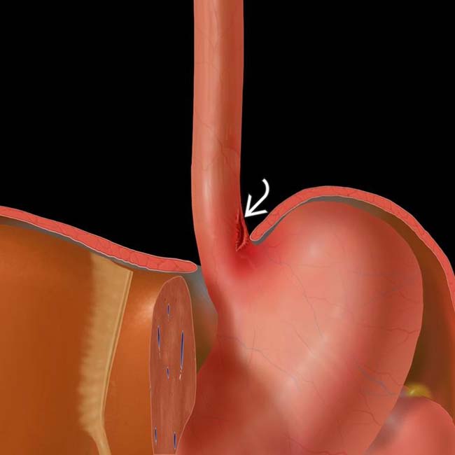
 of the distal esophagus, just above the hiatus and gastroesophageal (GE) junction.
 from a tear in the left anterior wall of the distal esophagus
  , a classic appearance for Boerhaave syndrome.
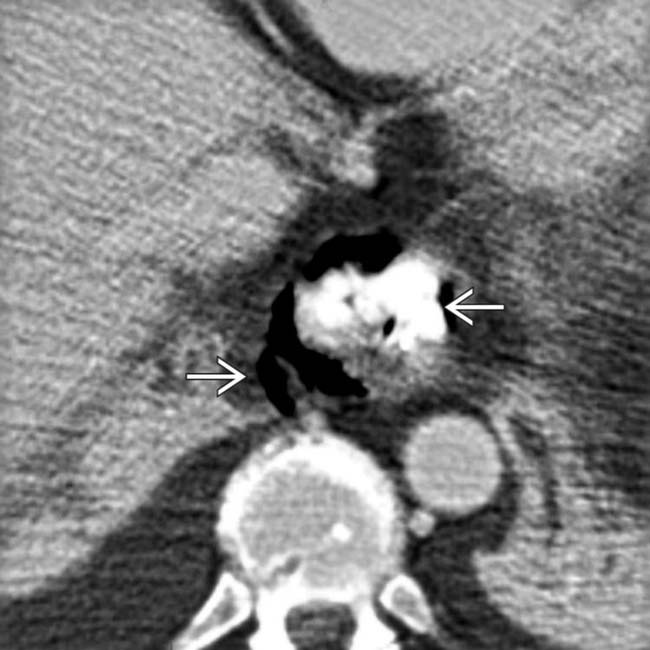
 surrounding the esophagus in the lower mediastinum and upper abdomen.
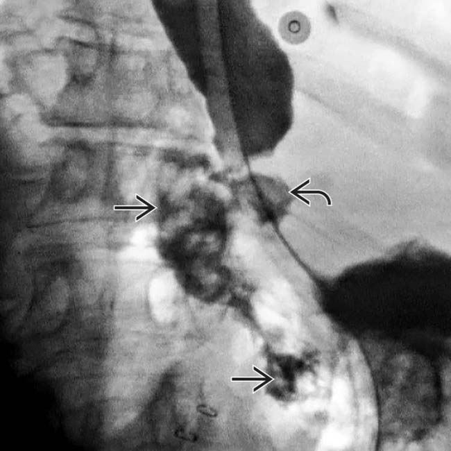
 . The site of the tear is the left anterior wall
  of the distal esophagus.
 Rarely from cervical or upper thoracic esophagus


