form an appropriate response to the pathology. Terms for aggressive periostitis include lamellated or “onion skin,” “hair-on-end,” “sunburst,” and Codman’s triangle.4
Table 29.1 PEDIATRIC BONE TUMORS BY PEAK AGE | |||||||||||||||||||||||||
|---|---|---|---|---|---|---|---|---|---|---|---|---|---|---|---|---|---|---|---|---|---|---|---|---|---|
|
Table 29.2 PEDIATRIC MULTIFOCAL BONE LESIONS | |||||||||
|---|---|---|---|---|---|---|---|---|---|
|
Table 29.3 PEDIATRIC LONG BONE TUMORS BY LOCATION | ||||||||||||||||||||||||
|---|---|---|---|---|---|---|---|---|---|---|---|---|---|---|---|---|---|---|---|---|---|---|---|---|
|
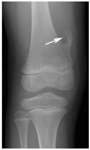
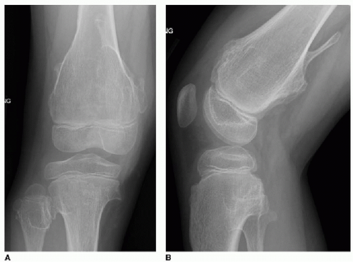
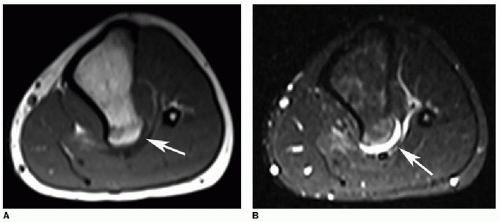
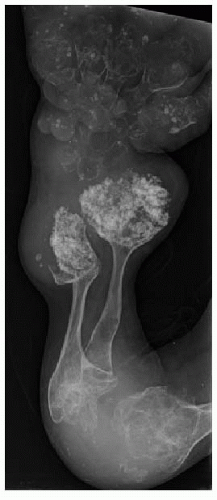
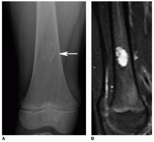
bone (Figs. 29.1 and 29.2). They most often arise from long bone metaphyses and are directed away from the joint. MR is helpful for identifying pathologic fractures and any overlying soft tissue abnormalities.1 On T2W or cartilage-sensitive sequences, the cartilaginous cap is normally a thin crescentic, high-signal structure (Fig. 29.3).
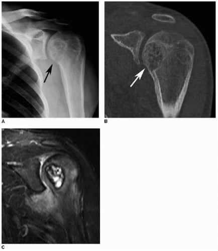
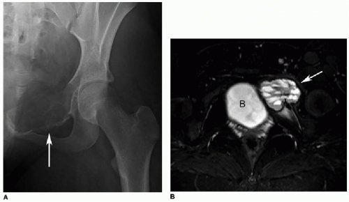
These tumors often present with pain and impaired function of the adjacent joint. Radiographs and CT will show a lobular lucent lesion with sclerotic margins and chondroid calcification, similar to an enchondroma (Fig. 29.7A, B). On MR, there is often surrounding marrow edema and an associated joint effusion (Fig. 29.7C). These peritumoral inflammatory changes are absent with enchondromas. Chondroblastoma is treated with curettage and bone grafting, with a 20% likelihood of local recurrence.1,8, 9 and 10
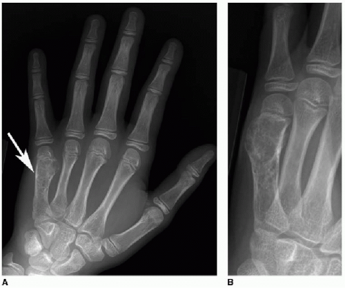
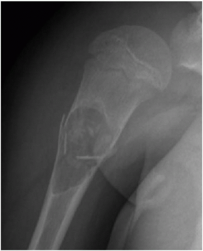
bone tumor or vascular malformation. In cases where a pre-existing lesion can be identified, the most common of these is giant cell tumor.12 ABCs are most common in adolescents and young adults and slightly more common in girls. They usually arise in long bone metaphyses and the posterior elements of the spine. Pain and swelling are typical presenting symptoms. As with SBC, treatment for ABC is curettage and bone grafting.1,12,13
appear as cortical-based ovoid lucencies with smooth sclerotic margins (Fig. 29.10). They tend to bulge toward the medullary cavity, and the surrounding cortex is intact. As fibroxanthomas involute, they become more sclerotic and less apparent on imaging. Treatment with curettage and bone graft is only indicated for larger lesions at risk for pathologic fracture.7,17, 18, 19 and 20
Stay updated, free articles. Join our Telegram channel

Full access? Get Clinical Tree


