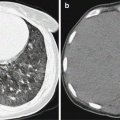
Fig. 3.1
Articular brucellosis. (a–d) At the late stage of the illness course, X-ray demonstrates narrowed interarticular space, sclerosis of articular surface, and bone hyperplasia at articular margin. In some cases, there are also bone destruction and articular effusion in a small quantity
3.7.1.3 CT Scanning
CT scanning demonstrates small multiple lesions of vertebral bone destruction, which are mostly confined at the vertebral margin. Around the lesions, obvious hyperplasia and sclerosis are demonstrated. In the newly formed bone tissues, new destruction lesions are mixed. The synovial cartilage or intervertebral disk is subject to destruction with equal density shadows. The joint surface is subject to hyperplasia and sclerosis, with increased density of adjacent bone and formation of paravertebral abscess in a small quantity. By contrast CT scanning, the margins of soft tissue and abscess are enhanced. At the early stage, the spine is demonstrated with small cystoid bone destruction at multiple vertebrae, with ring-shaped sclerosis at their margin. Such sclerosis mostly occurs at the superior and inferior margin of vertebra and the vertebral margin is subject to obvious osteoproliferation and osteosclerosis. At the late stage, the destruction area extends to the vertebral center, with compressed vertebra and wedge-shaped deformation of the vertebra (Figs. 3.2, 3.3, and 3.4).
Case Study 2
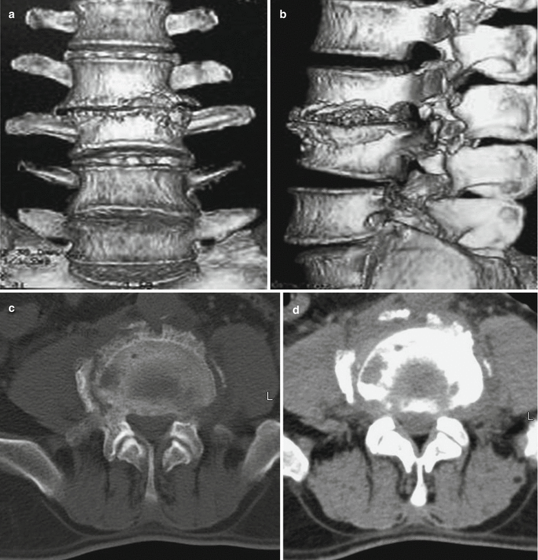
Fig. 3.2
Brucellar spondylitis. (a–d) CT scanning demonstrates osteoproliferation and bone destruction at the lumbar vertebrae, with narrowed intervertebral space. The vertebral margin is demonstrated with proliferation, hypertrophy, and ossification of the periosteum to form liplike or bank-like osteophytes. The newly formed osteophytes and the destructive lesions between them constitute characteristic lacelike vertebra. The osteophyte of adjacent vertebrae is demonstrated to connect with each other to form lateral-lateral vertebral fusion. Periostitis occurring at the transverse process is demonstrated as cap-like thickening at the top of the transverse process
Case Study 3
A male patient aged 24 years complained of lower back pain for 6 months that aggravated with accompanying numbness and pain at the right lower limbs for 1 month. He reported a history of working as a shepherd and lamb delivery. The serological agglutination test for Brucella was demonstrated positive.
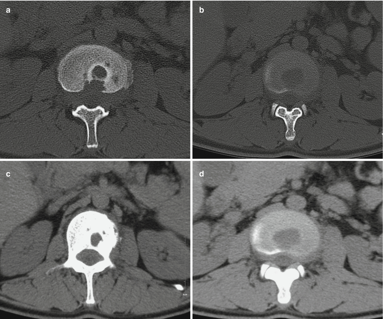

Fig. 3.3
Brucellar spondylitis. (a, b) Plain lumbar CT scanning demonstrates multiple bone destructions at the vertebra by bone window, with proliferation and sclerosis at the vertebral margin. The intervertebral disk is demonstrated with inner low-density lesions. (c, d) Soft tissue window demonstrates strips of low-density shadow around the diseased vertebra, which is actually formation of paravertebral abscess
Case Study 4
A male patient aged 47 years complained of lumbar pain, muscular pain at the lower limbs, and joint pain for 1 year. Serological agglutination test for Brucella demonstrated positive.
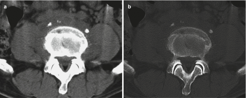

Fig. 3.4
Brucellar spondylitis. (a, b) Plain lumbar CT scanning demonstrates vertebral osteoproliferation, low uneven density shadow surrounding the vertebra with poorly defined boundaries. The vertebra is demonstrated with poorly defined interface to bilateral psoas majors. The psoas majors are demonstrated with slightly outward shift
(Note: Cases 3 and 4 as well as the corresponding figures were provided by Liu BL at the Second Affiliated Hospital, Harbin Medical University, Harbin, China.)
3.7.1.4 MR Imaging
Spinal margin bone destruction is the most common in the cases of brucellosis. The lesions are mostly multiple, invading vertebral margin of 1–2 vertebrae. In some rare cases, 3 vertebrae are involved. In the early stage, T1WI demonstrates low signal of different sizes, while T2WI, equal or high signal. After several weeks, bone defect lesions can be demonstrated as lower T1WI signal shadow, with no enhancement by contrast imaging. The lesions are demonstrated with irregular worm-bitten-like or sawlike appearance, with small surrounding abscess. In the cases of central spine type of brucellosis, the central lesions are demonstrated with rapid sclerosis, with no formation of deep bone destruction, no deformed vertebra due to compression. Vertebral proliferation and sclerosis are demonstrated by MR imaging as equal, high, or low signal. Early intervertebral disk involvement is demonstrated by T2WI and fat suppression imaging as increased signal at the intervertebral disk with different degrees, which are defined as edematous change, with no destructive effect on the vertebral end plate. Development of intervertebral disk involvement is demonstrated as thinner intervertebral disk, narrowed intervertebral space, and its connection to vertebral destruction, with irregular shape and formation of abscess. Paravertebral abscess is commonly demonstrated as long T1 long T2 signals. Contrast T1WI demonstrates marginal enhancement, with thick wall, well-defined boundary, and small abscess that do not exceed the length of diseased vertebra. There is no sign indicating downward flow of abscess fluid. The surrounding fat space is well defined. The MR imaging demonstrations of brucellosis at other joints are similar, commonly joint ossification, swelling of surrounding soft tissues, and articular effusion in a small quantity (Figs. 3.5, 3.6, 3.7, 3.8, and 3.9).
Case Study 5
A male patient aged 57 years was hospitalized due to lower back pain, accompanying muscular soreness and pain at both lower limbs and joint pain for more than 6 months.
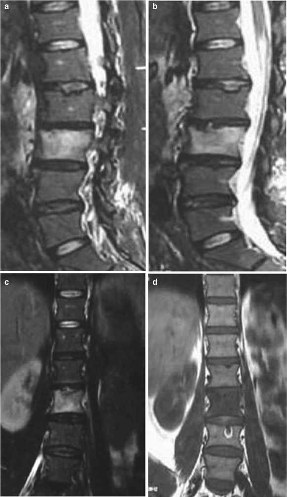
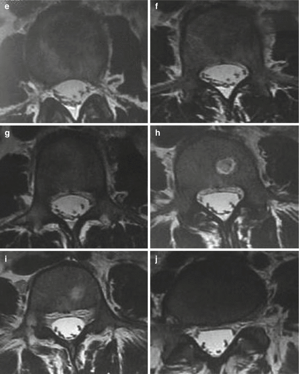


Fig. 3.5
Brucellar spondylitis. (a–j) MR imaging demonstrates patches of long T1 mixed slightly long T2 signals at the vertebrae of L2 and L3, with narrowed intervertebral space at the L2–L3, and strips of mixed slightly long T2 signal at the peripheral vertebra. Contrast imaging demonstrates abnormal signal with obviously uneven enhancement at the diseased vertebra and its periphery
Case Study 6
A male patient aged 51 years complained of intermittent lower back pain for more than 2 months.
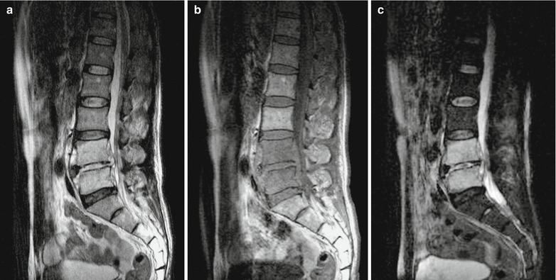

Fig. 3.6
Brucellar spondylitis. (a–c) MR imaging demonstrates slightly deformed L4–L5 with long T1 long T2 signals. By contrast imaging, the whole vertebra is demonstrated with homogeneous enhancement, and the intervertebral disk is demonstrated with uneven enhancement
Case Study 7
A male patient aged 64 years was hospitalized due to lower back pain, accompanying muscular soreness and pain at the lower limbs and joint pain for more than 6 months. He reported lower back pain with no known causes about half a year ago, with accompanying muscular pain at the lower limbs and major joint pain at the lower limbs. Recently, he received serological agglutination test for Brucella at the local CDC, with positive findings. And then he paid his clinic visit in our hospital. By laboratory tests, WBC count is 4.98 × 10⁹/L, GR % 64.1 %, ESR 50 mm/h, and CRP 10.8 mg/L. The clinical diagnosis was brucellar spondylitis. The symptoms disappeared after he received treatment for 2 months.
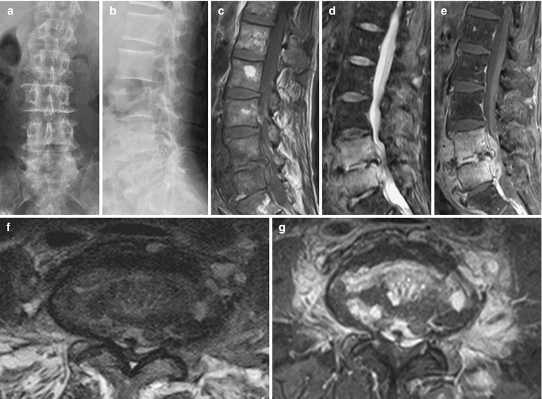

Fig. 3.7
Brucellar spondylitis. (a, b) Lateral and anterior-posterior imaging demonstrates intact sequence of lumbar vertebrae, obvious osteoproliferation at L2–L5 and bone destruction at the posterior margin of L5. (c, d, f) MR imaging demonstrates intact sequence of the lumbar vertebrae, diffuse slightly long T1 and long T2 signals at L4 and L5, no involvement of the appendices, obvious osteoproliferation at the L4 and L5, focalized destruction of the inferior end plate of the L4 and superior end plate of L5. The intervertebral space is demonstrated to be narrowed at L4–L5 and L5–S1, with internal long T2 signal and disk protrusion. Its posterior dural sac is demonstrated to be obviously compressed. And the paravertebral soft tissues are demonstrated with obvious swelling. The psoas major is involved. (e, g) Contrast imaging demonstrates obvious enhancement of the L4 and L5, irregular enhancement of intervertebral disk at L4–L5, obvious diffuse enhancement of the paravertebral soft tissues, enhancement of anterior and posterior longitudinal ligaments, and patches of enhancement at the left erector muscle of spine (Note: the case and figures were provided by the Department of Radiology, You’an Hospital, Beijing, China)
Case Study 8
A male patient aged 53 years was hospitalized due to intermittent fever and muscular pain for 23 days. He experienced fever 23 days ago, which was more severe after noons and during nights. He had the highest body temperature of 40 °C, with accompanying chills and aversion to cold. After clinic visit of a community-based clinic, he was diagnosed with viral infection and received medications for 4 days. His body temperature then returned to normal, but he began to experience muscular pain of the neck, with restricted cervical motions. Five days before his admission, the patient began to experience recurrence of fever with no known causes, with a body temperature of 38.7 °C and accompanying aversion to cold, chills, and lumbar paravertebral muscular pain. By physical examination after admission, the neurological system was normal, with normal brain nerves, muscle tension of limbs, and muscular strength. By bilateral finger-nose test and heel-knee-tibia test, he had favorable stability and accuracy. By bilateral needle stabbing, the sensation was symmetric. Tendon reflex of the limbs were symmetric and appropriate. No bilateral pathological signs were induced. The neck is soft; the Kernig test positive; and Patrick test positive. One week after the admission, the blood bacterial culture indicated growth of Brucella. Reexamination by standard tube agglutination test for Brucella is positive. The patient reported long-term intake of raw milk. The clinical diagnosis was brucellosis.
(For case detail and figures, please refer to Guo YJ et al. Chinese Journal of Contemporary Neurology, 2013, 13 (1): 49.)
Case Study 9
A female patient aged 42 years was hospitalized due to intermittent fever for 11 months, lower back pain for 7 months, and weakness of both lower limbs for 1 month. Her lower back pain is persistent. By physical examination, tenderness is near the spinous process of L3 and L4 and no reflex pain. By straight leg raising test, the right leg 70° and the left leg 90°, with muscular atrophy of both lower limbs. By laboratory test, WBC is 6.6 × 10⁹/L, and the antibody titer of Brucella was 1:16.
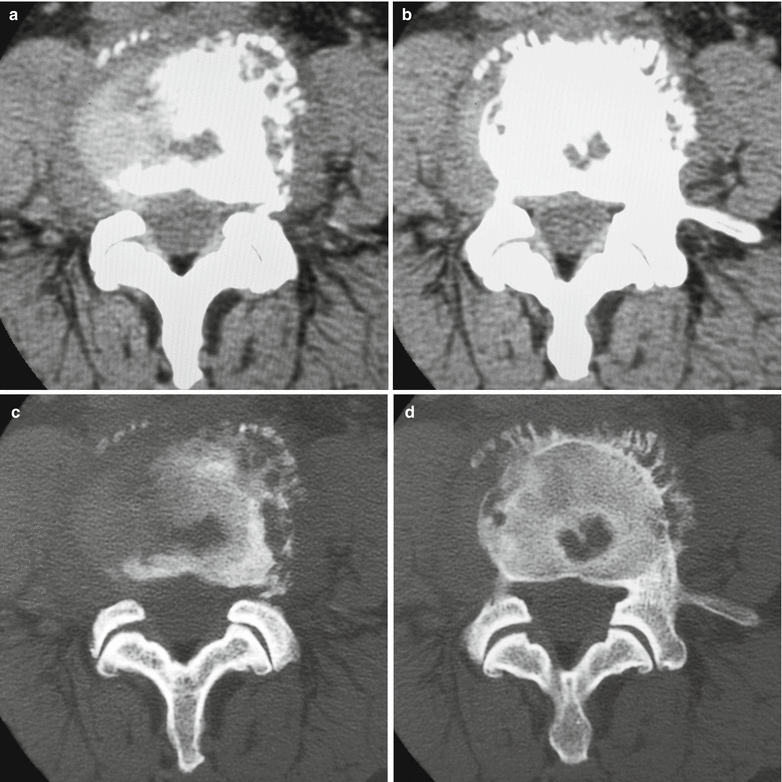
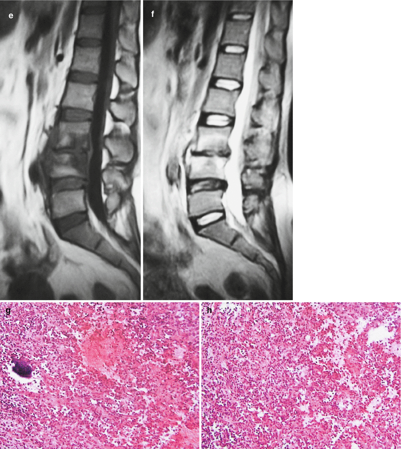


Fig. 3.8


Brucellar spondylitis. (a–d) CT scanning demonstrates multiple irregular bone destruction at the vertebral endplate of L4, with marginal sclerosis and no sequestrum. The bone destruction area is demonstrated to be surrounded by multiple lacelike osteophyte, with swelling of vertebral marginal soft tissue. (e, f) MR imaging demonstrates patches of long T1 and long T2 signals at the vertebrae of L3 and L4, with poorly defined boundaries. The corresponding intervertebral space is demonstrated to be narrowed, with swelling of paravertebral soft tissue and slightly compressed dural sac. (g, h) Pathology demonstrates chronic inflammation, formation of granuloma, fibrin-like exudation in a small quantity and accompanying small abscesses
Stay updated, free articles. Join our Telegram channel

Full access? Get Clinical Tree


