(Right) Double-contrast barium esophagram shows longitudinally oriented filling defects representing Candida plaques . This modality is quite accurate in depicting the characteristic mucosal plaques, ulcers, and less common manifestations of Candida esophagitis. However, in most patients, the combination of odynophagia and oral thrush is sufficient to make the diagnosis and begin treatment.
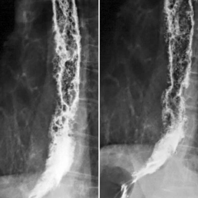
(Left) Esophagram shows a shaggy appearance of the esophagus due to ulcers and raised plaques. Note the innumerable pseudodiverticula , which are narrow, flask-shaped dilations of excretory ducts. These nonspecific findings have also been observed in patients with chronic esophagitis or dysmotility syndromes.
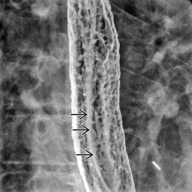
(Right) Esophagram shows a severely irregular surface pattern, due to innumerable plaques and ulcers. A mild stricture was present in the upper esophagus (not shown).
TERMINOLOGY
Synonyms
• Esophageal candidiasis, moniliasis
Definitions
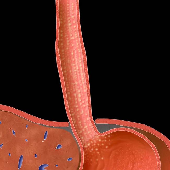
• Infectious esophagitis caused by fungi of Candida species, usually Candida albicans
IMAGING
General Features
• Best diagnostic clue
Mucosal plaques in immunocompromised patient
• Location
Any part or entire esophagus
Fluoroscopic Findings
• Double-contrast esophagram
Discrete plaques with longitudinal orientation
– Plaques are raised mucosal lesions (filling defects in barium pool); if there is central collection of barium, then lesion is an ulcer
“Cobblestone” or “snakeskin” appearance with confluent plaques
Severe cases: Deep ulcers
Only gold members can continue reading. Log In or Register to continue


 . This modality is quite accurate in depicting the characteristic mucosal plaques, ulcers, and less common manifestations of Candida esophagitis. However, in most patients, the combination of odynophagia and oral thrush is sufficient to make the diagnosis and begin treatment.
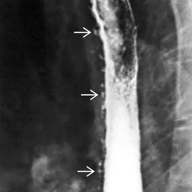
 , which are narrow, flask-shaped dilations of excretory ducts. These nonspecific findings have also been observed in patients with chronic esophagitis or dysmotility syndromes.