91 Cardiac Imaging: Myocardial Perfusion
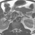
Figure 91.1 depicts a delay in perfusion of the inferior (or “posterior”) wall of the left ventricle at the midventricular level (arrows), on an image obtained in the short-axis orientation. Occlusions or constrictions within the arteries supplying blood to the cardiac muscle can lead to a reduction in or cessation of the flow of oxygenated blood reaching the myocardium, thereby inhibiting the supply of nutrients necessary for cellular activity and normal cardiac function. The MR approach used to assess the extent and uniformity of microvascular blood flow to the myocardium is referred to as cardiac perfusion imaging. Multislice, T1-weighted scans are acquired through the left ventricle within each cardiac cycle before, during, and after bolus administration of a contrast agent that decreases T1, typically a gadolinium chelate. The process provides a dynamic assessment of the rate and extent of blood with a short T1 (and thus high signal intensity) perfusing the left ventricle and can be analyzed both visually and with postprocessing software.
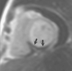
Fig. 91.1
Related posts:
Stay updated, free articles. Join our Telegram channel

Full access? Get Clinical Tree


