92 Cardiac Imaging: Myocardial Viability
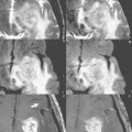
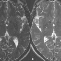
Myocardial muscle tissue that has been damaged by a reduction in the normal flow of oxygenated blood falls into three main categories. The first category, stunned myocardium, is living, viable tissue that has been subjected to short durations of reduced blood flow. In most cases, stunned myocardium will recover to normal function with time. The second category, hibernating myocardium, is that which has been subjected to long durations of reduced blood flow and requires interventional measures to restore normal cardiac function. The third category is necrotic or nonviable myocardium, tissue that no longer contains living myocardial cells or myocytes and is therefore unable to be returned to normal function. Patients with known cardiomyopathy resulting from decreased blood flow to the myocardium may benefit greatly from interventional measures if the area in question remains viable. However, if the tissue is no longer viable, there is little benefit in subjecting the patient to the possible risks of an interventional procedure. The differentiation between viable and nonviable myocardium is the focus of cardiac viability MR imaging. Figure 92.1 demonstrates a b-SSFP-based acquisition (see Case 27) where normal myocardium appears dark. The bright, enhanced myocardium in the apex (arrow) in this postinfarction image is thought to be nonviable due to the delayed collection of contrast in the tissue.
Related posts:
Stay updated, free articles. Join our Telegram channel

Full access? Get Clinical Tree


