Advances in cardiac gated computed tomography (CT) and magnetic resonance imaging (MRI) have significantly expanded the role of imaging in the evaluation of cardiac lesions. Echocardiography is the most common imaging study performed, and incidentally discovered cardiac lesions on echocardiography account for a significant proportion of cardiac mass evaluations. Additionally, the increasing temporal resolution of conventional CT allows better visualization of the heart on dedicated chest or abdominal examinations that may, in turn, increase the detection of cardiac masses.
Once a cardiac lesion is detected, dedicated cardiac imaging provides detailed information crucial for patient management. Anatomic localization of the lesion is the first and most important step in evaluating a suspected cardiac mass. To begin, a lesion must be confirmed to be of or from the heart. Occasionally, lesions mimicking masses turn out to be normal structures. Masses originating within the pericardium, mediastinum, and lung may invade the heart or compress the heart sufficiently to suggest falsely that they are cardiac masses. The epicardial fat is helpful in discerning whether a mass is truly cardiac in origin. Masses external to the heart displace this thin layer of fat toward the heart, whereas masses of the myocardium often outwardly displace or infiltrate this layer. Similarly, the pericardium can help in distinguishing an anterior mediastinal mass from a myocardial mass. Tenting of pericardium away from the heart or draping of the pericardium over the mass suggests a myocardial origin.
The location of a mass within the heart provides important diagnostic information and is the next step in evaluating myocardial lesions. Lesions originating from the valves are most commonly vegetations or adherent thrombus, although rare primary cardiac tumors such as papillary fibroelastoma may originate from the valves.
After localization, tissue characterization of cardiac lesions is paramount to diagnosis. For instance, detecting fat within a lesion narrows the differential diagnosis of a cardiac lesion considerably to include lipomas, teratomas, and lipomatous hypertrophy of the interatrial septum. Calcifications are rare in malignant myocardial tumors, with the exception of metastases that calcify, such as osteosarcoma and the extremely rare primary cardiac osteosarcoma. Cystic components are less helpful in differentiating myocardial tumors, although the presence of cystic components makes lymphoma less likely.
When a lesion is detected in the heart on imaging, regardless of the modality, the first clinical question is typically whether the mass is a thrombus or a neoplasm. Imaging findings favoring a neoplastic cause include vascularity and infiltration of the mass into the myocardium. Behavior on repeat imaging such as resolution with anticoagulation (which would indicate thrombus) is an important clinical diagnostic factor. Although contrast enhancement of a lesion excludes acute thrombus, organized thrombus may exhibit low levels of enhancement.
Finally, an understanding of the epidemiology of myocardial disease is helpful in organizing a differential diagnosis for cardiac lesions. Primary cardiac masses are rare, occurring with a frequency of between 0.002% and 0.19%. Most primary cardiac masses are benign, and more than half of benign cardiac masses are myxomas. Among malignant cardiac masses, metastatic disease is by far the most common, followed distantly by angiosarcoma and lymphoma.
Imaging of Cardiac Masses
Multiple imaging tools are employed in the evaluation of cardiac masses. Because each modality has strengths and weaknesses, echocardiography, cardiac CT, and cardiac MRI are often used in concert. Although transthoracic echocardiography is accessible and is often the first imaging modality used to assess cardiac masses, cardiac MRI has improved resolution and soft tissue contrast. Cardiac-gated CT can provide detailed anatomic information, can evaluate extent of local invasion, and can better assess calcium. Detailed anatomic and functional evaluation of myocardial masses is often required for surgical or radiation therapy planning. Additionally, postsurgical or follow-up examinations may be required. Understanding the available imaging modalities and their complementary nature is important when diagnosing a suspected cardiac mass or when recommending subsequent studies.
Radiography
Myocardial masses are rarely detected or evaluated using radiography. Even when large, intracavitary masses may not significantly distort the cardiac silhouette. When arising from the myocardium, masses alter the normal cardiac contour or appear as cardiac enlargement. Calcifications may be localized to the heart when they are present in masses such as myxomas. Masses in the left atrium may result in pulmonary venous congestion and pulmonary edema by impeding pulmonary blood flow back to the heart or obstructing the mitral valve.
Echocardiography
Ultrasound is a safe and effective means of evaluating both the anatomy and function of the heart. Echocardiography is the most common imaging study, and imaging evaluation of heart disease most often begins with this modality. Cardiac masses are frequently first detected as an incidental finding on echocardiography. Multiple imaging planes are available, thus allowing targeted examination of the cardiac chambers, the cardiac valves, the pericardium, and the proximal aorta. Real-time imaging provides valuable information about myocardial global and regional wall motion. Doppler echocardiography allows estimation of important functional parameters such as ejection fraction, stroke volume, and cardiac output. Valvular dysfunction can also be quantified. Serial imaging with echocardiography provides important surveillance information for worsening cardiac dysfunction. Newer echocardiographic tools such as three-dimensional echocardiography have been developed.
Because ultrasound is so frequently used in the evaluation of heart disease, cardiac masses are often first discovered by this modality. Masses in the fetal heart such as rhabdomyoma are almost always first discovered during fetal ultrasound, and important mimics of cardiac masses such as thrombus or vegetations are often first characterized by echocardiography. Mass characterization on echocardiography often centers on vascularity, attachment site and morphology (e.g., stalklike or broad-based attachment site), and relationship with adjacent anatomy. Transesophageal echocardiography is the best modality to evaluate small masses arising from the cardiac valves. Real-time evaluation can depict valvular dysfunction associated with a cardiac mass, such as prolapse of a pedunculated atrial myxoma across the mitral or tricuspid valve.
Echocardiography is operator dependent and targeted, and certain regions of the heart and cardiac masses are challenging to detect. The left ventricular apex, inferior vena cava, and aortic arch may be difficult to image. Small masses centered in the myocardium may be visible only as focal thickening of the myocardium. Distinguishing myocardial from pericardial origin may be impossible with large and infiltrating masses. Additionally, the extent of extracardiac disease is not well evaluated with echocardiography, particularly into the pulmonary and systemic veins. Adjacent lungs, airways, and calcifications can be obstacles to detailed mass characterization when using echocardiography. Body habitus can limit transthoracic echocardiography.
Computed Tomography
Multidetector CT has established a role in imaging cardiac tumors, primarily secondary to its short imaging time, its high spatial resolution, and its unsurpassed ability to evaluate calcification. Depending on imaging parameters such as heart rate and mode of imaging, the entire heart can be imaged in as little as one heartbeat. Submillimeter detector arrays provide spatial resolution superior to that of MRI. Additionally, high spatial resolution in three planes (isovoxel) during image acquisition allows for post hoc multiplanar reconstructions. CT is the best imaging modality to demonstrate calcifications. The presence of calcification within a mass may provide important diagnostic information, and linear calcification in the myocardium indicative of earlier myocardial infarction can provide important information when thrombus is suspected. Fat can also be readily identified on CT as low attenuation regions (−30 to −100 Hounsfield units) within cardiac masses.
Fundamental to the use of CT to evaluate cardiac tumors is electrocardiogram (ECG) gating. Not only does it eliminate blurring secondary to cardiac motion, but also it can provide functional analysis. If scanned retrospectively such that the heart is effectively imaged throughout the entire cardiac cycle, an intracavitary mass can be depicted in both diastolic and systolic positions. This is perhaps most applicable when a mass is associated with or originating from a cardiac valve. Cardiac movement may reveal the pedunculated nature of the mass or assist in evaluating for the site of origin or attachment. Finally, high spatial resolution of cardiac CT when combined with ECG gating is an advantage of this modality over MRI.
Intravenous, nonionic contrast material is an important element of evaluating cardiac masses on CT. Opacification of the cardiac chamber helps to delineate the borders of an intracavitary mass. Contrast may also depict the differential enhancement of normal myocardium from an intramyocardial mass. The timing and composition of the contrast bolus are special considerations when performing cardiac CT. If the approximate anatomic location is known before imaging, the contrast bolus can be timed to opacify the involved or most adjacent cardiac chamber when the images are acquired. This typically involves bolus tracking or test bolus techniques, in which an anatomic site such as the ascending aorta is serially imaged in rapid succession after contrast injection to trigger the scan (bolus tracking technique) or to time the diagnostic contrast bolus (test bolus technique). Configuring the contrast bolus is particularly challenging when evaluating a right atrial mass. Inflow of nonopacified contrast material from the inferior vena cava and coronary sinus and concentrated contrast material inflowing from the superior vena cava and causing beam-hardening artifact may obscure the borders of a mass. Accurate imaging may require injecting a dilute mix of contrast material and saline solution after the typical concentrated contrast bolus, or early delayed scans (90 seconds) after injection of a large amount of contrast material to capture the second phase after systemic venous return. Injecting contrast material from a lower extremity vein may be considered when evaluating masses involving or near the inferior cavoatrial junction or when a mass has obstructed the superior vena cava.
When considering cardiac CT for the evaluation of myocardial masses, the examiner must be familiar with the limitations of this modality. Significant and persistent arrhythmias can make adequate ECG gating difficult or impossible. Similarly, high resting heart rates may limit the spatial resolution secondary to excessive motion artifact. Temporal resolution is lower in CT as compared with both echocardiography and MRI. With the exception of calcification, tissue characterization is inferior to that available with MRI. CT uses ionizing radiation, which is potentially hazardous. This factor is particularly important to consider when imaging young patients. Finally, cardiac CT almost always employs the use of intravenous contrast material, which is potentially nephrotoxic.
Magnetic Resonance Imaging
The high tissue contrast resolution, multiplanar imaging capability, and standardized and reproducible imaging planes make MRI the modality of choice for imaging most cardiac tumors. Unlike MRI examinations of other anatomic structures that may rely on standardized protocols and imaging planes, cardiac MRI for myocardial masses often requires real-time input from the cardiac imager to characterize a mass optimally and to delineate its anatomy more clearly. Anatomic definition and tissue characterization are evaluated using T1-weighted, proton density, and T2-weighted sequences. Cystic components and hemorrhage products can be distinguished using these sequences as elsewhere in the body. Gradient echo cine images provide wall motion and functional data and may demonstrate the dynamic interaction of a mass with cardiac valves. Early T1-based sequences and delayed enhancement sequences after gadolinium administration are usually employed both to evaluate enhancement and vascularity of the mass and to detect underlying myocardial scars when thrombus is suspected.
Imaging protocols for evaluating cardiac tumors on MRI usually begin with scout images and fast spin echo axial images, which provide early localization of the tumor. From this point, two-chamber steady-state free precession (SSFP) sequences are prescribed, depending on the location of the tumor (e.g., oriented along the tricuspid valve and right ventricular apex for tumors in the right side of the heart, oriented along the mitral valve and left ventricular apex for tumors in the left side of the heart). Four-chamber and short-axis SSFP sequences are then acquired, to provide an anatomic overview of the heart and functional information. T2-weighted triple-inversion recovery sequences are used to evaluate edema or necrosis in the mass and adjacent myocardium.
Gadolinium is an important component of the MRI evaluation of cardiac masses and should be used when possible. The principles of gadolinium enhancement were originally developed to evaluate myocardial infarction. T1-weighted fast spin echo sequences are acquired before and after the administration of intravenous gadolinium to detect abnormal enhancement of the mass or to highlight the differential enhancement compared with normal myocardium. Cardiac masses may enhance after the administration of intravenous gadolinium by one mechanism or by a combination of several mechanisms. Gadolinium accumulates in the extracellular space. When cellular damage has occurred, such as in necrotic or fibrotic portions of a mass, gadolinium may enter the intracellular space and result in enhancement. Another mechanism of gadolinium enhancement is its collection in a region of increased extracellular space, as in the case of myocardial edema. Local myocardial hyperemia incited by the presence of a mass may increase delivery of gadolinium to the mass.
Cardiac MRI has a role not only in tumor characterization but also in differentiating mass from thrombus, an important and common clinical question when a mass is discovered. MRI has superior soft tissue resolution and better reproducibility than echocardiography. MRI is a valuable tool for both the diagnosis and follow-up imaging of intracavitary thrombus. Intravenous gadolinium is also helpful in differentiating tumor from thrombus in that thrombus is typically avascular and therefore not expected to enhance. Large, chronic thrombus may demonstrate thin peripheral enhancement near the attachment site from neovascular ingrowth.
MRI is also useful for determination of the degree of myocardial infiltration and pericardial involvement. Differential enhancement of tumor and normal myocardium may provide important information about infiltrating tumors. Localized thickening of the pericardium in association with a cardiac mass may suggest involvement of the pericardium. An enhancing pericardial nodule or the presence of a hemopericardium in the setting of a cardiac tumor provides compelling evidence of the malignant nature of the cardiac tumor and indicates neoplastic pericardial involvement.
Limitations of MRI include long imaging times, the presence of contraindications such as claustrophobia or preexisting pacemakers, accessibility, and poor spatial resolution. Calcification is not well evaluated on MRI, an important component of tissue characterization best depicted on CT. Nephrogenic systemic fibrosis, previously known as nephrogenic fibrosing dermopathy, is a disease characterized by fibrosis of the skin and internal organs that has been associated with gadolinium. This association is derived from gadolinium found in biopsied fibrotic tissues in patients with this disease. All patients with nephrogenic systemic fibrosis have a history of renal insufficiency (except one report of two transplant recipients in which the donors’ exposure to gadolinium was not reported). Relatively poor spatial resolution of cardiac MRI compared with echocardiography and CT limits its evaluation of very small masses, especially when associated with cardiac valves.
Benign Cardiac Masses
Myxoma
Background
Comprising approximately 50% of primary cardiac neoplasms, myxoma is the most common primary cardiac tumor. Histologically, myxomas are mesenchymal in origin, although they are distinct from noncardiac myxomas. Most sporadic myxomas occur in adults between 30 and 60 years old. Cardiac myxomas may also be seen in younger patients with Carney complex, an autosomal dominant syndrome of cardiac myxomas, hyperpigmented skin lesions, and extracardiac neoplasms such as breast fibroadenomas, pituitary adenomas, and psammomatous melanotic schwannomas. The myxomas in the setting of Carney complex may be uncharacteristically multifocal, extraatrial, and recurrent.
Most (>90%) cardiac myxomas are solitary, atrial, and intracavitary. They tend to arise from the interatrial septum (78%) near the fossa ovalis. From this site, they are more commonly within the left atrium (~80%) than the right atrium, and they may occasionally grow through the fossa ovalis to occupy both atria. Approximately 16% of these tumors contain calcification at surgical resection. Myxomas may be cystic, necrotic, or hemorrhagic. Although most are broad based, their occasional stalklike attachment to the cardiac chamber wall is a distinctive feature when present.
The clinical presentation of cardiac myxoma is attributable to embolization, functional obstruction, or idiopathic symptoms. Systemic and central nervous system embolization from these tumors may be from bland thrombus associated with the mass or from tumor embolization. Even embolization into the coronary arteries that causes small myocardial infarction has been described. When intracavitary and atrial, these masses may clinically mimic mitral or tricuspid stenosis.
Imaging
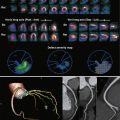
The imaging appearance of cardiac myxomas reflects the pathologic features, although internal structure characterization is nonspecific ( Fig. 32-1 ). Although most myxomas are homogenous on echocardiography, they may show internal hypoechoic structures consistent with cysts or hyperechoic foci if calcifications are present. These masses are heterogeneously low attenuating on cardiac CT. CT is the optimal modality to evaluate for calcification and may show heterogeneity reflecting hemorrhage or necrosis. In the absence of calcification or hemorrhage, myxomas typically have high signal intensity on T2-weighted imaging because of their high water content, are isointense to myocardium on T1-weighted images, and demonstrate heterogeneous low-grade enhancement with gadolinium administration. Features that confer greater confidence in making the diagnosis of myxoma include stalklike attachment to the interatrial septum and mobility. In addition, prolapse of an atrial mass through the mitral or tricuspid valve is also frequently seen in myxomas and can be demonstrated on echocardiography, CT, and MRI. However, prolapse through the tricuspid valve is more frequently encountered when tumor thrombus extends from the IVC into the right atrium such as from renal, liver, adrenal, retroperitoneal, or uterine tumors. Tumor prolapse through the mitral valve may also occasionally be encountered by lung masses that invade the atrium through the pulmonary veins. Hence, if a prolapsing tumor is encountered, it is critical to identify the entire mass and its origin and attachment site.

Cardiac Papillary Fibroelastoma
Background
Cardiac papillary fibroelastoma is a connective tissue tumor (cardiac endothelial papilloma) lined by a single layer of endothelium. It is the second most common benign primary cardiac tumor based on large surgical and pathologic series, and it represents 10% of all primary cardiac tumors. Papillary fibroelastomas are by far the most common tumors to involve the cardiac valves, with greater than 75% of these tumors occurring in this location. The aortic valve is the most common valve affected (29%), followed by the mitral and tricuspid valves. The mean age at diagnosis is approximately 60 years, with a slight male predominance.
Papillary fibroelastomas are typically less than 1 cm in size, although tumors ranging up to 7 cm have been reported. They often have a stalklike attachment to the avascular valvular endocardium. They tend to be on the aortic side of the aortic cusps and on the atrial side of the atrioventricular valves. That these tumors usually do not arise from the free leaflet edges or coaptation margins may help differentiate them from vegetations. The central portion and villous fronds of connective tissue are also avascular, and the overall appearance has been compared to a sea anemone on gross pathologic examination. Elastic fiber layers are the hallmark of these tumors when present in histologic samples.
Most patients with cardiac papillary fibroelastomas are asymptomatic, and most tumors are discovered incidentally. The friable nature of these tumors, the associated thrombus, and their predilection for the valves account for their clinical presentation in symptomatic patients. Stroke and transient ischemic attacks are the most frequent presenting symptoms, and peripheral embolization has also been reported. Despite the predilection of these tumors for the cardiac valves, valvular dysfunction is not typical.
Imaging
Cardiac papillary fibroelastomas are most frequently discovered on echocardiography ( Fig. 32-2 ). Classic echocardiographic findings include a small, pedunculated mass with peripheral stippled edges, attributed to the echogenic interfaces of small papillary projections. Advances in CT and MRI have established a role for these modalities in the evaluation of this tumor.

Surgical excision is curative and is therefore the treatment of choice for symptomatic cardiac papillary fibroelastomas. Tumor mobility is an independent predictor of major morbidity and mortality. Surgery may also be considered in an asymptomatic patient if the mass is mobile.
Lipoma
Background
Cardiac lipomas are encapsulated tumors composed of adipose tissue, typically arising from a broad pedicle extending from the epicardium. These tumors are rare, although multiple intramyocardial cardiac lipomas have been associated with tuberous sclerosis. They may arise from the epicardium, myocardium, or interatrial septum.
Symptomatic, cardiac lipomas have been known to cause cardiac arrhythmias, obstruction when intracavitary, and symptoms such as shortness of breath secondary to local mass effect. Lipomatous hypertrophy of the interatrial septum is a distinct entity that is discussed separately later. Differentiating features are its absence of a capsule and the finding that this tumor is composed of immature brown fat.
Imaging
Cardiac lipomas are typically incidental findings, seen on chest radiographs as an abnormal contour of the cardiomediastinal silhouette or on echocardiography as a homogenous, hyperechoic mass when intracavitary. The echocardiographic appearance of lipomas in the pericardial space is more varied, and tumors in this location may be heterogeneous, hyperechoic, or hypoechoic. Necrosis and hemorrhage are uncommon. CT and MRI provide superior tissue characterization ( Fig. 32-3 ). Lipomas on CT are homogeneous, fat attenuation, encapsulated masses without demonstrable enhancement. On MRI, these masses are T1 hyperintense and suppress with chemical fat saturation. These pliable masses may encase the coronary arteries without obstruction. Septations, if present, are thin and without nodularity or a nonfatty component.