, Valery Kornienko2 and Igor Pronin2
(1)
N.N. Blockhin Russian Cancer Research Center, Moscow, Russia
(2)
N.N. Burdenko National Scientific and Practical Center for Neurosurgery, Moscow, Russia
Cavernous angiomas, cavernous malformations, were allocated into the group of true malformations in the international histological classification of tumors of the central nervous system only in 1979. Before this, cavernous angiomas (CAs) were regarded as tumors. CAs represent the system of communicating vascular cavities with various sizes and sinusoid shapes, filled with blood. Vascular cavities are separated by connective partitions with different thicknesses, which are common to several adjacent microcavities. The walls of the cavities are lined with the endothelium forming papillary excrescences. Each cavity is characterized by an independent argyrophil framework, lack of muscle, and elastic fibers; capillaries or intertwined bands of endothelial cells can be located between them. There is no brain tissue in the CA structure; however, there can be cysts, areas of thrombosed masses, sclerosis, and calcificates. The origin of sclerotic changes in the majority of cases is unequivocal—development of clots characteristic of CA. Microhemorrhages within the malformation, as well as beyond its borders, are considered a typical feature of CAs. The perifocal area is characterized by reactive changes in the glia with its yellow discoloration due to imbibition of the brain substance with hemosiderin accumulated in macrophages. The size of CAs varies from several microns to several centimeters. In a hemorrhage inside the cavernoma, a rupture of the inter-cavity walls and formation of larger cavities are possible. This results in the fact that in some cases cavernous angioma consists of only a few cavities filled with blood, i.e., they are essentially a one- or multi-chamber hematoma.
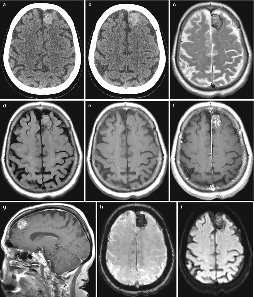
Diagnosis of cavernous angioma on the background of hematoma on CT and MRI is very difficult, which is due to several factors—a small size of the malformation and a possible self-destruction of the malformation in case of the hemorrhage.
MR manifestations of hematomas are more specific. In the early stages, contrast enhancement of hematomas is determined primarily by the presence of water molecules—they look isointense in T1-weighted images and hyperintense in T2-weighted images. In the following hours, oxyhemoglobin transforms to deoxyhemoglobin, which substantially shortens the relaxation time T2 in the hematoma area. The latter becomes very dark in T2-weighted images but is still isointense on T1-weighted MRI. Further oxidation results in the formation of methemoglobin, which significantly increases the signal from hematoma in T1-weighted and T2-weighted images. An increase in the MR signal (especially on T1-weighted MRI) extends from the periphery to the center, indicating lysis of erythrocytes and “release” of methemoglobin—intracellular methemoglobin has a high MR signal in T1-weighted images and a low signal in T2-weighted images, and “free” methemoglobin is characterized by a high MR signal in all sequences (Figs. 34.1 and 34.2). At the late subacute phase and early chronic phase, on the periphery of the hematoma, a narrow area of a low signal, visible in T2-weighted images, begins to form, represented by the deposition of hemosiderin in macrophages of the hematoma capsule. By this time, the hematoma has an increased signal from the center and a low signal as a thin rim from the periphery in all sequences (Fig. 34.3).