Cerebral Venous Thrombosis

Chapter 14 Cerebral Venous Thrombosis


Cerebral venous thrombosis (CVT) was first described by Ribes in the early 19th century on the basis of postmortem examination.1 For a long time, CVT was seen as a rare and severe illness resulting in seizures, focal deficits, and often death. Its association with sinus infections was well described,2 but other predisposing conditions were not yet recognized. However, advances in vascular neuroimaging have led to a renewed appreciation of this disorder, which is more common, more variable, and less uniformly severe than previously assumed.36


Cerebral sinus venous thrombosis is estimated to account for 0.5% of all strokes in adults,3 but its true incidence is unknown, and frequent underrecognition is suspected.7 Initially asymptomatic venous sinus thrombosis has been implicated in the etiology of idiopathic intracranial hypertension.8,9 Indeed, studies have demonstrated that the most common symptom of CVT is headache (80%–90%) and the most common sign papilledema (50%–60%).10 Other clinical signs include focal deficits and partial seizures, and alteration of consciousness. Headache may be the only clinical symptom of CVT.11,12 Approximately one quarter of all patients have completely normal examination.3


Multiple causes and risk factors for CVT have been identified.5 The list comprises genetic and acquired prothrombotic conditions (the latter most prominently including pregnancy, puerperium, and combination of hormonal contraceptives and smoking), infections (particularly sinusitis, otitis, mastoiditis, and meningitis), systemic inflammatory illnesses (such as systemic lupus erythematosus, sarcoidosis, and inflammatory bowel disease), hematological disorders (e.g., polycythemia, leukemia, thrombocythemia, paroxysmal nocturnal hemoglobinuria), systemic cancer, severe dehydration, and head trauma. The threshold for obtaining brain imaging to exclude CVT should be lower in the presence of these conditions.


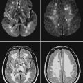
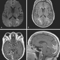
Current neuroimaging techniques have greatly enhanced our ability to diagnose CVT.3,13 Magnetic resonance imaging (MRI), particularly in combination with MR venography (MRV), provides excellent diagnostic yield in cases of thrombosis of dural sinus or deep cerebral veins (Figure 14-1).13,14 It is worth reemphasizing that noninvasive imaging modalities are allowing us to learn the broad spectrum of CVT. Although once the diagnosis was only suspected after severe intracranial hypertension or venous infarctions had occurred, today the possibility of CVT must be considered in the differential diagnosis of patients with new headaches and benign intracranial hypertension. Often CVT can be timely diagnosed only by keeping a low threshold for its clinical suspicion. A delayed or missed diagnosis of CVT, unfortunately still a common occurrence in practice, cannot be justified now that we have reliable and extremely safe means to reach the diagnosis.
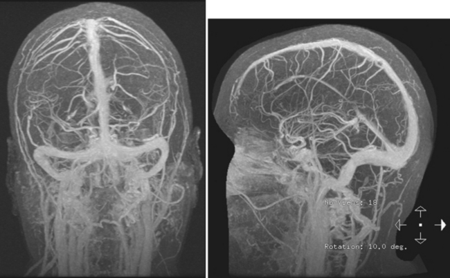


Although MRI/MRV is the most proved and widely used set of tests for the identification of CVT, computed tomography (CT)/CT venogram (CTV) represent a valuable alternative when MRI is contraindicated or unavailable. The main disadvantage of CTV is the requirement for administration of iodinated contrast material.


Case Vignette


A 23-year-old woman presented during her puerperium for evaluation of new severe headaches. They had developed soon after child delivery and had worsened over the previous several days. She described them as bifrontal and pulsating. They were not accompanied by nausea or vomiting, but photophobia was intense. Soon after initial evaluation, the patient experienced increasing vomiting and developed right facial weakness and double vision on right lateral gaze. She did not have any significant medical history except for several early miscarriages and heavy smoking. She had recently begun using an oral hormonal contraceptive.


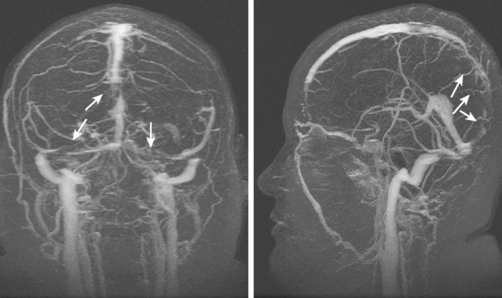
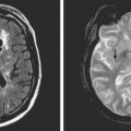
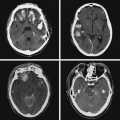
On examination at the hospital, the patient was drowsy, had bilateral papilledema, right VI nerve palsy, and peripheral right facial nerve palsy. Otherwise the examination was unremarkable. She underwent MRI and MRV of the brain that demonstrated extensive thrombosis of the posterior two thirds of the superior sagittal sinus and the proximal transverse sinuses (Figure 14-2). Thrombophilia workup was negative.


The patient improved gradually after initiation of anticoagulation. Anticoagulation was continued for 1 year, and she was advised to avoid contraceptives and stop smoking.


Knowledge of the normal anatomy of the cerebral venous system and its variations is essential to interpret the vascular images effectively. Hence we begin by illustrating and summarizing basic anatomical information.


ANATOMY OF THE VENOUS SYSTEM


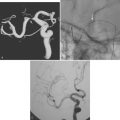
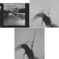
The cerebral venous system consists of dural venous sinuses, superficial veins, and deep veins. The dural venous sinuses are venous channels devoid of valves situated between the two layers of the dura, and thus they are not collapsible. They constitute the major draining pathways of the cerebral venous circulation. Cerebral veins are also devoid of valvular structures and have a very thin wall with no muscular tissue. They empty into the dural sinuses but may reverse their flow in cases of dural sinus occlusion. Venous anatomy is illustrated on angiographic pictures shown in Figure 14-3.


Dural Sinuses


The major dural sinuses are the superior and inferior sagittal sinuses; cavernous and intercavernous sinuses; superior and inferior petrosal sinuses; occipital sinus; and straight, transverse, and sigmoid sinuses.


The major superficial sinus is the superior sagittal sinus, which is located in the convex margin of the falx cerebri. It runs from the foramen cecum to the torcula Herophili, where it merges with the transverse sinuses. It receives blood from cortical parasagittal veins on the cortical surface.


The inferior sagittal sinus is much smaller and is contained in the posterior half or two thirds of the free margin of falx cerebri. It terminates at the falcotentorial apex by joining with the great cerebral vein (vein of Galen) to form straight sinus. It receives blood from the falx, corpus callosum, cingulum, and medial cerebral hemispheres. The occlusion of this sinus is rarely clinically significant.


The straight sinus is situated at the line of the junction of the falx cerebri with the tentorium cerebelli. After a descending course, it terminates at the internal occipital protuberance, usually emptying into the left transverse sinus. It receives venous blood flow from the great vein of Galen and superior cerebellar veins, thus participating in the deep venous drainage. Occlusion of this sinus usually produces venous infarcts in the deep basal ganglia.


The transverse sinuses are contained within the attachment of the tentorial leaves to the calvarium. At the posterior border of the petrous temporal bone, the transverse sinuses receive the superior petrosal sinus to become the sigmoid sinuses. Transverse sinuses receive blood from the superior sagittal sinus and straight sinus, as well as bridging veins from cerebellum, inferolateral surfaces of the temporal and occipital lobes, and tentorium. It also receives blood from the cortical vein of Labbé. In more than half of cases, the right transverse sinus is larger than the left.15,16 In up to 20% of the cases, a narrowed or atretic segment can be identified in at least one of the transverse sinuses.16


The sigmoid sinuses begin where the transverse sinus leaves the tentorial margin and drain into the jugular bulb, thus becoming the internal jugular veins. There are numerous anastomoses between sigmoid sinuses and the vertebral plexus, muscular, and scalp veins.


The superior petrosal sinuses extend from the cavernous sinus to the sigmoid sinus, running along the attachment of the tentorium to the dorsal ridge of the petrous temporal bones. They receive venous drainage from the pons and upper medulla, lateral mesencephalic vein, cerebellar veins, and veins draining the inner ear.


The inferior petrosal sinuses lie in a groove between the petrous apex and the clivus, extending posterolaterally along the petrooccipital fissure. They have multiple anatomical variations but usually terminate draining into the jugular bulb. On their way, they form multiple anastomoses with deep venous plexuses.


The cavernous sinuses lie on either side of the sphenoid body. They are formed by numerous small veins with multiple interconnections that constitute a trabeculated space. They extend from the superior orbital fissure to the petrous apex. They contain the cavernous segment of internal carotid artery and cranial nerve VI. Cranial nerves III, IV, and V1 branch of trigeminal nerve are located in the lateral wall of the sinus. This sinus receives venous drainage from superior and inferior ophthalmic veins, intercavernous sinus, and multiple deep venous plexuses. They drain into the superior and inferior petrous sinuses.


Arachnoid granulations are present along venous sinuses and often protrude into their lumen. They can be confused with areas of restricted flow on imaging studies when they are prominent (most frequently the case in the transverse and superior sagittal sinus).


Deep Cerebral Veins


Deep venous drainage is variable, although major basal veins are relatively constant. The deep venous system drains the inferior frontal lobe; most of the white matter of the frontal, temporal, and parietal lobes; basal ganglia; thalamus; corpus callosum; and upper brainstem. Small medullary veins are responsible for draining blood from the deep cerebral white matter and basal ganglia into the subependymal veins of the lateral ventricles. Those veins form the thalamostriate veins, which unite with septal veins to form the internal cerebral veins. The internal cerebral veins are the largest deep veins. They are paired and originate behind the foramen of Monro. After collecting venous drainage from deep white matter and basal ganglia, they join to form the vein of Galen.


The basal veins of Rosenthal arise deep within the Sylvian fissure. They course posteriorly around the cerebral peduncles and across the tectum to join the vein of Galen. The anterior and deep middle cerebral veins as well as veins from the insula and cerebral peduncles drain through this system.


The great cerebral vein of Galen arises under the splenium of the corpus callosum from the junction of the internal cerebral veins and basal veins of Rosenthal. It usually runs a short trajectory before joining with inferior sagittal sinus to form the straight sinus.


This complex anatomical composition can be simplified by knowing the major venous drainage territories. Four general pathological patterns can be identified in cases of CVT:


IMAGING CHARACTERISTICS OF CVT


Computed Tomography


Signs of Venous Thrombosis


Noncontrast CT scan may disclose the delta sign (Figure 14-4), a dense triangle that represents an acute thrombus at the end of the superior sagittal sinus or torcula. However, this sign is insensitive and lacks specificity. It is present in less than a quarter of cases of superior sagittal sinus thrombosis17 and may be mimicked by blood hyperviscosity (e.g., dehydration, erythrocytosis), sinus calcification, or adjacent subarachnoid or subdural hemorrhage.13

Contrast CT scan may demonstrate the empty delta sign (Figure 14-5), caused by lack of contrasted flow in the occluded sinus but enhancement of the sinus wall. It is slightly more sensitive than the delta sign and may be present in close to 30% of cases of superior sagittal sinus thrombosis.17 This sensitivity may be substantially greater when thin sections of helical CT scan are carefully reviewed.13 The empty delta sign is probably produced by rich dural venous collateral circulation (from meningeal venous tributaries) and a vascular mesh of cavernous spaces within the dural wall.17


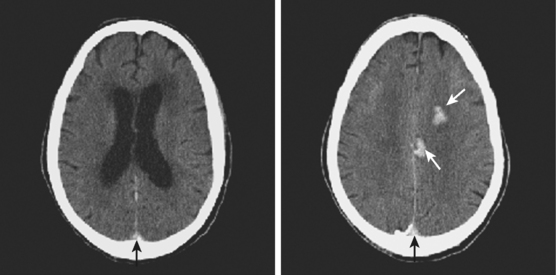
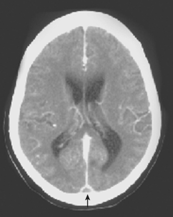
Signs of Parenchymal Involvement


< div class='tao-gold-member'>

Stay updated, free articles. Join our Telegram channel

Jul 23, 2016 | Posted by in NEUROLOGICAL IMAGING | Comments Off on Cerebral Venous Thrombosis

Full access? Get Clinical Tree

Get Clinical Tree app for offline access