60 Cervical Plexus Block
The cervical plexus derives from the ventral rami of C2, C3, and C4. Its superficial branches primarily consist of the lesser occipital, great auricular, transverse cervical, and supraclavicular nerves. These nerves innervate the skin of the neck and shoulder. The nerves converge at the posterolateral border of the sternocleidomastoid muscle, where cervical plexus block can be performed. Common indications for cervical plexus block include carotid endarterectomy, thyroid and parathyroid surgery, and surgery on the clavicle.1
Ultrasound imaging is useful for guiding blocks of the cervical plexus. Sonographic landmarks can be identified and in some cases direct nerve imaging is possible. Optimal needle tip placement with in-plane approach can be used to target injections and potentially avoid complications.2 Cervical plexus blocks are sometimes classified as superficial or deep according to whether or not the nerves have pierced the prevertebral fascia at the site of injection. Intermediate cervical plexus blocks target the cervical plexus where its nerves lie underneath the sternocleidomastoid muscle.3 This technique is highly amenable to ultrasound guidance and is described here.
Suggested Technique
Several sonographic landmarks more precisely determine the level for cervical plexus block injections (Table 60-1):
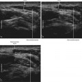
1. The great auricular nerve is the largest branch of the cervical plexus and is often visible as it lies over the sternocleidomastoid muscle. Cervical plexus block can be performed just caudal to the point at which the great auricular nerve wraps around the posterolateral edge of the sternocleidomastoid muscle to join the remainder of the cervical plexus.
2. The external jugular vein crosses the posterolateral edge of the sternocleidomastoid muscle. The cervical plexus emerges at the muscle edge to lie underneath the external jugular vein. This vein can serve as a tubular conduit to promote distribution of the injection to the underlying cervical plexus.
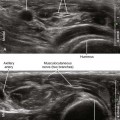
3. Cervical levels can be estimated by cervical vertebral morphology on ultrasound imaging scans.4,5 The anterior tubercle of the seventh cervical transverse process is rudimentary. In contrast, the anterior tubercle of the sixth cervical transverse process (sometimes referred to as Chassaignac’s tubercle) is prominent compared with its posterior tubercle. This bony morphology can be used to identify higher cervical levels when performing cervical plexus blocks. In addition, the first segment of the vertebral artery lies anterior to the seventh cervical transverse process before it enters the cervical spinal canal. The intervening space (sulcus) between the anterior and posterior tubercles becomes progressively smaller as one slides the transducer cephalad. This is because the exiting ventral rami are smaller at the higher cervical vertebrae. The cervical plexus can be blocked just deep to the sternocleidomastoid muscle at the level of the C4 foramina as assessed in a transverse plane of imaging.
4. At the level for cervical plexus block, the ventral rami of the brachial plexus are not visible and the scalene muscles taper off to small diameter. At this level the longus capitis and levator scapulae muscles are more prominent.
Table 60-1 Sonographic Landmarks for Intermediate Cervical Plexus Block*
| Location for Intermediate Cervical Plexus Block | Comment on the Sonographic Landmarks |
|---|---|
| Where the EJ crosses the posterolateral edge of the SCM | The cervical plexus emerges to lie underneath the EJ. |
| Where the GAN rounds the posterolateral edge of the SCM | The GAN is the largest branch of the cervical plexus and therefore relatively easy to identify. |
| At the level of the C4 transverse process | |
| Where the brachial plexus and scalene muscles are no longer visible | The cervical plexus emerges between the longus capitis and levator scapulae muscles. |
EJ, External jugular vein; GAN, great auricular nerve; SCM, sternocleidomastoid muscle.
* This block is performed with a transverse view of the posterolateral edge of the sternocleidomastoid to guide injection underneath this muscle where the cervical plexus organizes. The level of block is chosen to help complete distribution of the injection to the entire cervical plexus. The transducer slides in a cephalocaudad fashion to identify these landmarks for intermediate cervical plexus block. Although the quality of these sonographic landmarks can vary from patient to patient, all four locations are very close to each other
Related posts:


Stay updated, free articles. Join our Telegram channel

Full access? Get Clinical Tree


