44.1 Lateral XR: normal
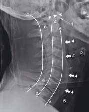
The cervical vertebrae are visible down to T1. The anterior (A), posterior (B) and spinolaminar (C) spinal lines are aligned normally. There is no injury of the bones or prevertebral soft-tissue (4). The atlanto-axial distance is normal (arrowheads)
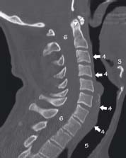
CT appearances of the same patient as in fig 44.1. Normal alignment is shown with no evidence of bone or soft tissue injury. Here the atlanto-axial space (arrowheads) is seen clearly
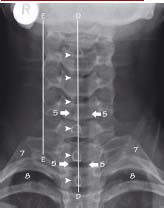
The spinous processes (line D) are normally aligned and equally spaced (arrowheads). The lateral alignment (line E) is also normal on both sides
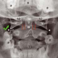
The alignment of the lateral masses of C1 and C2 (*) is normal. The spaces between the peg (2) and the lateral masses of C1 (1) is also equal (red lines). The peg itself is partly obscured by the occipital bone (10)
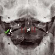
The spaces between the peg (2) and lateral masses of C1 (1) are not equal (red lines). The peg (2) which is an anterior structure does not align with the spinous process (11) which is a posterior structure. The head is rotated with respect to the X-ray beam. Most importantly, however, the lateral masses still show good alignment (*)
key
1 Atlas (C1)
2 Odontoid peg of axis (C2)
3 Hyoid bone
4 Prevertebral soft tissues
5 Trachea
6 Spinal canal (contains spinal cord)
7 First rib
8 Lung apex
9 Teeth
10 Occipital bone
11 Spinous process
Lines
A Anterior spinal line
B Posterior spinal line
C Spinolaminar line
D Spinous process line
E Lateral margin line
Related posts:


Stay updated, free articles. Join our Telegram channel

Full access? Get Clinical Tree


