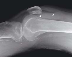
A vertical split fracture is seen on the lateral side of the tibial plateau (arrowheads)
25.2 Tibial plateau fracture: lateral knee
The fracture is not easily seen on this view but a fat-blood interface is seen (arrowheads). This is known as a lipohaemarthrosis (fat and blood in a joint)
25.3 Ankle fracture
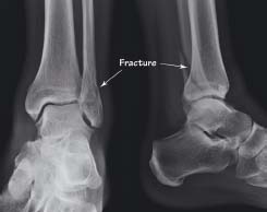
There is an oblique fracture of the lateral malleolus (distal fibula). This is at the level of the ankle joint and can therefore be classified as a Weber B type fracture
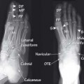
25.4 Lisfranc injury
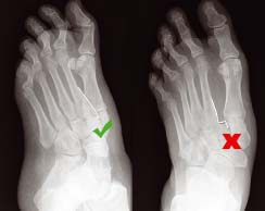
This is an example of how some injuries are only visible on one view. The DP (dorsiplantar) view (right) shows loss of alignment of the medial edges of the second metatarsal and the middle cuneiform. Alignment appears normal on the oblique view (left)
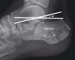
There is flattening of the calcaneus with reduction of Bohler’s angle (*) to 15° (normally 20-40°). Multiple fractures involving the subtalar joint were caused by falling from height and landing on the heels. The patient also had spinal injuries – a common combination
25.6 Osteoarthritis
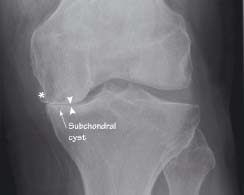
The knee is a common site for osteoarthritis. Here there is loss of the medial joint space (arrowheads) with articular surface sclerosis (increased density of bone) and formation of subchondral cysts. A large marginal osteophyte (*) is also present. These are the four cardinal features of osteoarthritis
Tibial plateau fractures
These fractures are often complex and include vertical split and depression fractures. The full extent of injury is frequently difficult to appreciate on plain X-ray imaging and requires further imaging, for example with CT, before planning surgery. Plain X-ray imaging may demonstrate a lipohaemarthrosis. Fractures of the lateral plateau are the most common, associated with a high impact force, and have the worst prognosis. They are usually caused by impaction of the lateral femoral condyle on the tibial plateau.
Lipohaemarthrosis
Related posts:


Stay updated, free articles. Join our Telegram channel

Full access? Get Clinical Tree


