45.1 Odontoid peg fracture: peg view
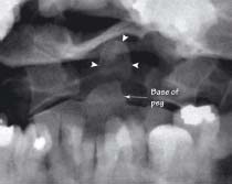
A fracture is seen through the base of the odontoid peg. The tip of the peg is displaced (arrowheads). This is a type 2 odontoid peg fracture
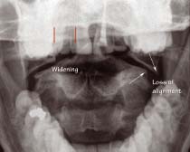
There is widening in the gap between the odontoid peg and the lateral masses of the atlas (red lines). The lateral edges of the atlas/C1 and axis/C2 (arrows) are no longer aligned due to a burst Jefferson-type fracture. Compare with normal alignment (chapter 44)
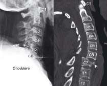
This patient presented with severe lower neck pain and tenderness after a fall downstairs. The lateral XR does not show down to the T1 vertebral body despite several attempts. This is because the shoulders are obscuring the view. A CT of the C-spine (right) revealed a fracture at C7 with malalignment of all three spinal lines
45.4 Rheumatoid arthritis (sagittal T2 MR image)
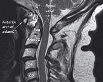
(Same patient as figs 45.5 and 45.6.) In this patient with chronic rheumatoid arthritis, neck pain and neurological symptoms, there is thick high signal material anterior to the peg. This pannus (synovial thickening) has eroded the peg and destroyed ligaments supporting the atlanto-axial joint, leading to subluxation of this joint (see figs 45.5 and 45.6). This has also caused narrowing of the spinal canal at this level
45.5 Lateral C-spine: extension view
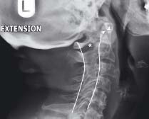
(Same patient as figs 45.4 and 45.6.) In this position the odontoid peg is shown to be eroded but is closely applied to the anterior arch of the atlas (arrowheads). There is plenty of space in the spinal canal (*) for the spinal cord
Related posts:


Stay updated, free articles. Join our Telegram channel

Full access? Get Clinical Tree


