Challenge

Chapter 3 Challenge


Comment


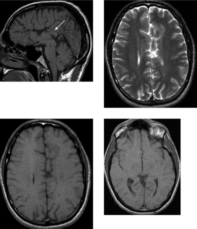
The HPEs are a spectrum of disorders characterized by hypoplasia of the rostral end of the neural tube and the premaxillary segment of the face (lack of forebrain induction). HPE is characterized by failure of cleavage of the embryonic prosencephalon, which is normally complete by embryonic day 35. With this comes partial to complete failure of separation of the telencephalon and diencephalon into the right and left cerebral hemispheres and basal ganglia or thalami, respectively. Because the optic vesicles and olfactory bulbs evaginate from the prosencephalon, visual disturbances and incomplete formation of the olfactory system are frequently present. Hypoplasia of the premaxillary segment results in facial anomalies, including cleft lip and palate; abnormalities of the orbit (cyclopia, hypotelorism); and forehead proboscis.


Holoprosencephaly may be divided into subtypes: alobar (the most severe form), semilobar, lobar (the mildest form of the major subtypes), and middle interhemispheric variant. There is no clear distinction between subtypes. In alobar HPE, the falx, interhemispheric fissure, and septum pellucidum are absent. There is failure of separation of the cerebrum and the ventricular system, resulting in a monoventricle contiguous with a dorsal cyst. In semilobar HPE, the interhemispheric fissure and falx cerebri are usually formed posteriorly and absent anteriorly. In lobar HPE, the interhemispheric fissure and falx anteriorly are hypoplastic. Often the posterior corpus callosum or splenium is formed, as in this case (arrow). The third ventricle is usually well formed. In semilobar and lobar HPE, the ventricular system shows variable degrees of development. The middle interhemispheric variant differs from classic HPE in that the posterior frontal and parietal lobes are most significantly affected rather than the basal forebrain. The anterior frontal lobes and occipital lobes are separated, and the genu and splenium of the corpus callosum are formed, but the callosal body is absent. The hypothalamus and lentiform nuclei appear separated, but the caudate nuclei and thalami are incompletely separated in many cases. Heterotopias and cortical dysplasia are common associated anomalies of HPE, as seen in this case, in the left greater than right medial frontal lobes.


CASE 152 Olivopontocerebellar Degeneration


CASE 153 Band Heterotopia—Pachygyria


Comment


Heterotopias are migrational abnormalities in which normal neurons occur in abnormal locations as a result of failure of migration along the radial glial fibers from the germinal region to the cortex. Three well-recognized types of heterotopia include focal, subependymal, and diffuse (laminar, band). Subcortical heterotopia is a newer term that refers to a specific entity in which neurons are abnormally located predominantly in the subcortical white matter. The cortical dysplasias are different from the heterotopias in that they do not represent a failure of normal migration, but rather a failure in the development of a normal six-layered cortex after migration from the germinal matrix to the cortical region.


Diffuse or band heterotopia refers to a layer of gray matter whose migration has arrested such that it is localized between the subcortical white matter laterally and the deep white matter medially. The inner and outer margins of the heterotopic neurons are well demarcated. Therefore, the band of gray matter is separated from the overlying cortex by a mantle of subcortical white matter. Band heterotopias are frequently associated with overlying cortical dysplasias (pachygyria, polymicrogyria), as in this case. The severity of the cortical dysplasia is directly related to the severity of the band heterotopia: the thicker the band, the more severe the overlying dysplasia will be. Similarly, patients with band heterotopia tend to have more pronounced clinical symptoms. In addition to seizures, which may have an age of onset ranging from infancy to young adulthood, many children have moderate to severe developmental delay.


Hemimegalencephaly refers to hamartomatous overgrowth of a cerebral hemisphere. Migrational abnormalities are usually present within the affected hemisphere, but they may also be present on the contralateral side. On imaging, there is usually enlargement of the affected cerebral hemisphere and the ipsilateral lateral ventricle, as well as associated cortical dysplasias.


CASE 154 Huntington’s Disease


Comment


A variety of disease processes affect the extrapyramidal nuclei (basal ganglia, thalami) as well as the nuclei in the brainstem. These conditions are most commonly degenerative or metabolic, and many are inherited. Among the neurodegenerative processes that can affect the deep gray matter are Huntington’s disease, Wilson’s disease, and Hallervorden-Spatz syndrome. Toxic exposures may also result in abnormalities of the deep gray matter. Lesions in these structures typically result in movement disorders that can occur in isolation or in combination and include the following subtypes: abnormalities in muscle tone, involuntary movements, abnormal postural reflexes, and the inability to carry out voluntary movements. Toxic exposures that affect the basal ganglia include carbon monoxide, cyanide, hydrogen sulfide, ethylene glycol, and toluene. Although there is wide variation in the deep gray matter structures involved, many diseases have characteristic involvement of specific structures.


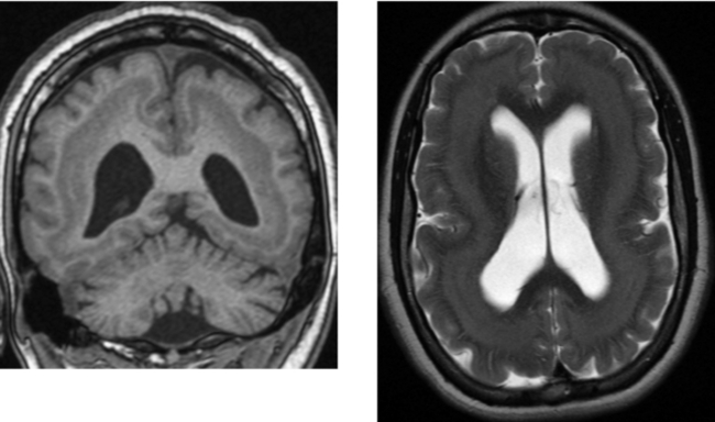
Huntington’s disease is inherited in an autosomal dominant manner that results in progressive degeneration of brain cells. The gene responsible for the disease is located on chromosome 4. Clinical manifestations typically include involuntary movement (choreoathetosis), rigidity, dementia, and emotional instability. Cognitive deficits are believed to be due to abnormal connectivity between the deep gray matter and the cortex. The disease typically presents in the fourth or fifth decade of life. The disease is progressive, with death occurring 15 to 20 years after its onset. On imaging, as in this case, Huntington’s disease is characterized by atrophy of the caudate nuclei, which results in ballooning of the frontal horns of the lateral ventricles (“boxcar” ventricles). There may also be involvement of the putamen, which can atrophy. MR imaging shows signal changes in these nuclei. These changes may be hyperintense on T2W images, as in this case, which is believed to be related to gliosis; other cases show T2W hypointensity, which is likely related to iron deposition. Other major imaging findings include cortical atrophy.


CASE 155 Meningocele and Pseudomeningocele of the Skull Base (Petrous Apex)


Comment


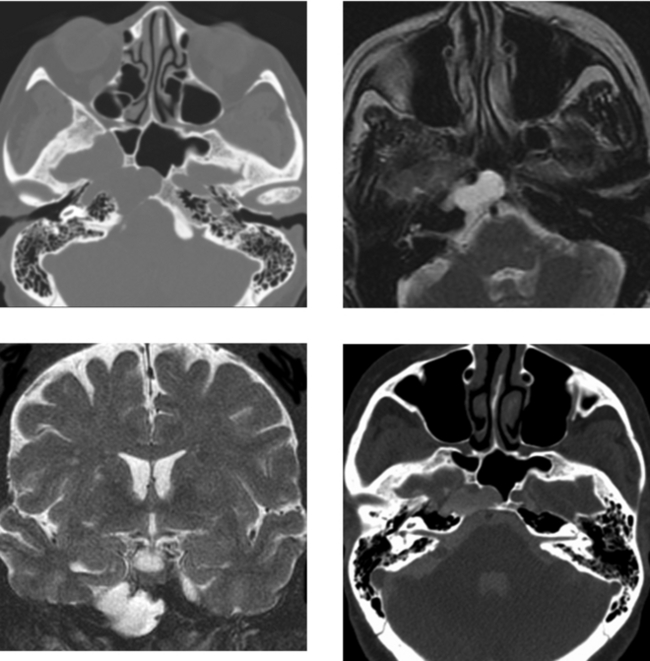
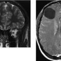
This case shows a well-demarcated, chronic-appearing lesion of the right petrous apex. There is expansion, smooth cortical bone thinning without bone destruction, and cortication medially, suggesting a long-standing process. The lesion is isointense to CSF on T2W images, and on the coronal T2W image, it is difficult to determine whether this lesion is separate from or an extension of Meckel’s cave into the osseous base of the skull. CT cisternography (instillation of contrast material into the thecal sac by lumbar puncture, followed by CT imaging after graceful tilting of the patient so that the contrast agent spreads through the subarachnoid spaces in the head) confirms that the petrous apex lesion communicates with the CSF spaces.


The differential diagnosis of a benign-appearing, expansile petrous apex mass includes cholesterol granuloma, mucocele, epidermoid, meningocele, and occasionally, an aneurysm of the internal carotid artery. Mucoceles are frequently unilocular and show peripheral enhancement. The signal characteristics of mucoceles will vary, depending on the protein concentration and viscosity. Epidermoid cysts may have a benign appearance, or they can have more concerning radiologic findings, such as bone erosion or destruction. They typically appear similar to CSF on T1W and T2W images and hyperintense on FLAIR imaging, and demonstrate restricted diffusion. Cholesterol granulomas, when large, may be multilocular. There is usually thinning of the cortex with large lesions without bone destruction. Cholesterol granulomas are often hyperintense on all pulse sequences. In particular, the marked hyperintensity on unenhanced T1W images distinguishes these from many other lesions, and the presence of hemorrhage–fluid levels within these lesions is highly characteristic.


CASE 156 Right Thalamic Glioblastoma


Comment


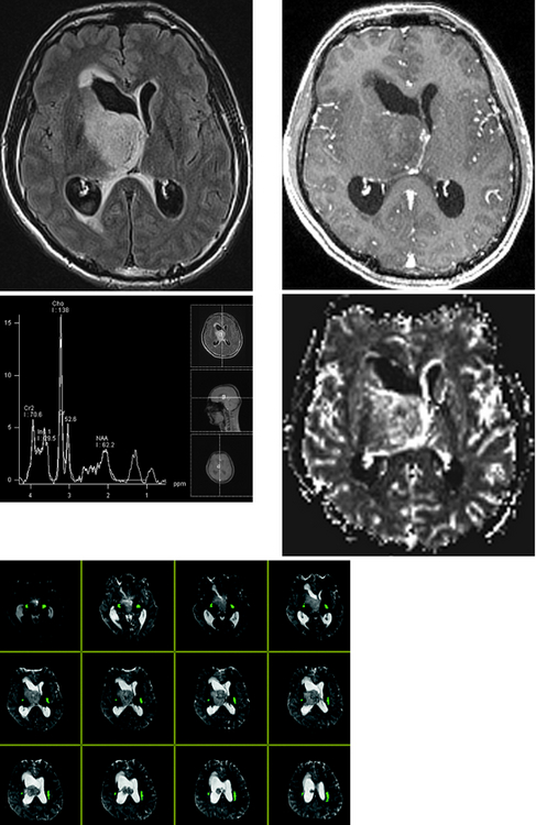
This case illustrates a solid mass centered in the right thalamus, with an exophytic component extending into the ventricle. There is mass effect, with partial obstruction of the foramen of Monroe resulting in hydrocephalus and transependymal flow of CSF along the margin of the right lateral ventricle. Most GBMs enhance and usually demonstrate heterogeneity because of the presence of necrosis or hemorrhage. Margins of the enhancing component are usually ill defined, and there is irregular thick peripheral enhancement due to central necrosis. Enhancement may extend into the adjacent white matter. In a newly identified brain tumor in which biopsy is anticipated, regions of enhancement correlate with regions of solid tumor on pathologic examination. Therefore, contrast is useful in identifying areas for stereotactic biopsy. In addition, enhanced images may identify tumor spread to regions that otherwise would not be noticed on unenhanced images, such as the leptomeninges, subarachnoid space, or subependymal region along the ventricular margins.


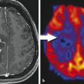
In this less typical case, the GBM is centered in the deep gray matter (most GBMs occur in the cerebral lobes), has no significant central necrosis, and shows only mild central enhancement. The functional in vivo techniques of MR spectroscopy and MR perfusion imaging allow correlation of metabolic activity with the vascular properties, respectively, and provide further insight into the grade of the primary glial neoplasm. In this case, these techniques were helpful in characterizing the high-grade nature of this neoplasm. Specifically, proton spectroscopy shows an elevated choline:NAA ratio (>2), and perfusion imaging shows markedly elevated rCBV, indicative of a high-grade neoplasm. For the neurosurgeon planning stereotactic biopsy, location of eloquent areas of brain relative to the tumor is critical to reduce potential neurologic deficits. Diffusion tensor imaging tractography in this case clearly identifies the corticospinal tracts (green).


CASE 157 Multiple Sclerosis and Glioblastoma


Comment


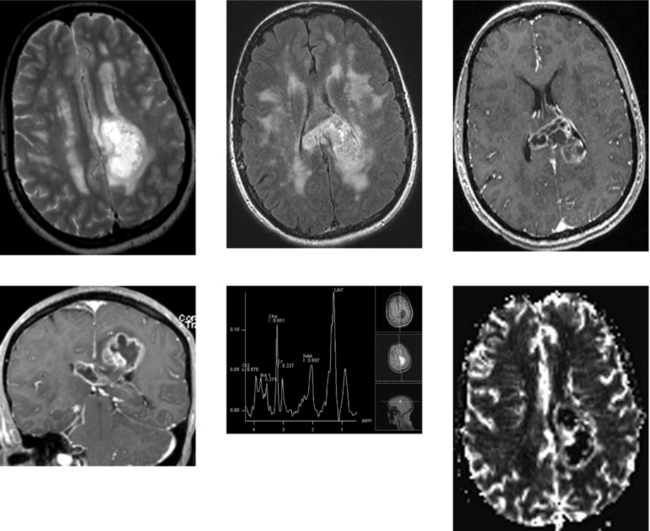
“Tumefactive” multiple sclerosis, high-grade glioma (GBM), and occasionally an abscess can appear similar on imaging, particularly in the absence of a clinical history. Multiple sclerosis typically occurs in younger patients, and there are often additional clinical or imaging findings to suggest this diagnosis. On close questioning, patients often have neurologic symptoms that are spaced both in time and in location. Furthermore, MR imaging may demonstrate white matter lesions separate from the mass that are suggestive of multiple sclerosis.


Advanced MR imaging sequences may be of value. Perfusion imaging in this case shows markedly elevated regional cerebral blood volume in the enhancing rim of the necrotic mass, suggesting a high-grade glioma or GBM rather than demyelination. GBM was confirmed at surgical biopsy. Nonspecific spectroscopic findings in tumefactive multiple sclerosis include elevation of choline, lactate, and lipid peaks, and a decrease in the N-acetylaspartate peak. These spectroscopic characteristics reflect the histologic correlate of marked demyelination in the absence of significant inflammation. Gliomas also consistently show reductions in N-acetylaspartate and increases in phospholipids, reflecting the replacement of normal neuronal tissue with a proliferating cellular process. Increases in lactate are not uncommon as a result of tissue ischemia and necrosis, as in this case. Variable increases in lipid levels may be seen.


Elevation of glutamate or glutamine peaks favors tumefactive demyelination and is typically not seen in aggressive neoplasms (not present in this case). Serial proton MR spectroscopy can be a useful, noninvasive method of overcoming the diagnostic dilemma of differentiating glioma from acute tumefactive demyelination. Persistent elevation of choline and lactate levels favors a glioma. Normalization of the initial increases in phospholipids, lipid, and lactate peaks within 3 to 4 weeks, followed by persistent, marked reductions of the neuronal marker N-acetylaspartate, has been described over time in tumefactive demyelination.


CASE 158 Adrenoleukodystrophy


Comment


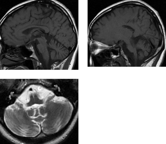
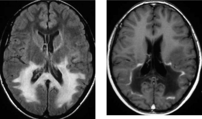
Adrenoleukodystrophy is an X-linked or autosomal recessive disorder that is related to a single enzyme deficiency (acyl coenzyme A synthetase) within intracellular peroxisomes. This enzyme is necessary for β oxidation in the breakdown of very long-chain fatty acids that accumulate in erythrocytes, plasma, and fibroblasts, as well as the CNS white matter and adrenal cortex. Boys typically present between the ages of 4 and 10 years. The clinical presentation may include behavioral disturbance, visual symptoms, hearing loss, seizures, and eventually spastic quadriparesis. Patients often present with adrenal insufficiency (Addison’s disease), which may occur before or after the development of neurologic symptoms.


As in other demyelinating and dysmyelinating disorders, MR is the imaging modality of choice for the detection of white matter disease, being far superior to CT. In adrenoleukodystrophy, the most common pattern of white matter disease is bilaterally symmetric abnormalities within the parietal and occipital white matter, extending across the splenium of the corpus callosum. The disease may continue to progress anteriorly to involve the frontal and temporal lobes. The region of active demyelination, usually along the anterior margin, may show contrast enhancement. Less typical presentations include predominantly frontal lobe involvement or holohemispheric involvement. Adrenoleukodystrophy also involves the cerebellum, spinal cord, and peripheral nervous system. Findings on MR imaging correlate well with neuropsychological measures.


CASE 159 Medulloblastoma


Comment


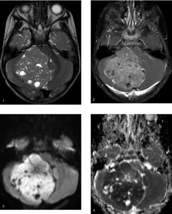
Medulloblastomas account for up to one third of all pediatric posterior fossa tumors. They occur more commonly in boys than in girls (approximately 3:1) and arise from the superior medullary velum of the fourth ventricle from primitive neuroectoderm. In children, they are typically midline masses associated with the inferior vermis, but occasionally may present as a lateral cerebellar hemispheric mass, as in this case. Importantly, subarachnoid seeding of the leptomeninges is very common at presentation (reported in up to 30% of cases in some series); therefore, patients should have a screening contrast enhanced MR imaging study of the spine to exclude this type of spread.


On unenhanced CT, medulloblastomas are typically hyperdense relative to brain parenchyma because of their dense cellularity. They are demarcated masses, and calcification, cystic change, or hemorrhage may be present in up to 10% to 20% of lesions. On MR imaging, the signal characteristics of medulloblastomas vary considerably on T2W imaging, depending on the presence of hemorrhage and the degree of cellularity. Most medulloblastomas show avid but heterogeneous contrast enhancement. Typically, they efface the fourth ventricle and present with hydrocephalus.


Accurate preoperative diagnosis is important in pediatric cerebellar tumors because this may affect the surgical approach. Diffusion MR imaging allows assessment of microscopic water diffusion within tissues, and with neoplasms, this diffusion seems to be primarily based on cellularity. Increasing cellularity leads to increased signal intensity on diffusion imaging and hypointensity on corresponding apparent diffusion coefficient (ADC) maps. Studies have suggested that diffusion imaging and ADC values may be useful in helping to distinguish among histologic types of pediatric brain tumors. Juvenile pilocytic astrocytomas have shown high ADC values and ratios. In contrast, medulloblastomas that characteristically are cellular have shown restricted diffusion, as in this case, with high signal intensity on diffusion images (image 3) and hypointensity on ADC maps (image 4). The cellularity of ependymomas is between that of astrocytomas and that of medulloblastomas. Hence, pilocytic astrocytomas are most often hyperintense on ADC maps, medulloblastomas are hypointense, and ependymomas fall somewhere in between.


CASE 160 Middle Cerebral Artery Territory Stroke Mimicking a Bone Metastasis


Comment


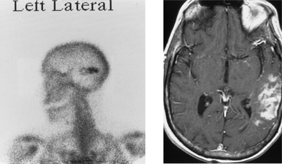
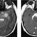
The left lateral projection from the patient’s delayed bone scan shows increased radiotracer activity projecting over the temporal bone region (it is important to remember that an anteroposterior view is necessary to accurately localize the abnormality). This was the only abnormality on the patient’s bone scan. Corresponding enhanced MR imaging shows mild local mass effect in the superficial portion of the posterior left temporal lobe with cortical enhancement; the imaging findings are consistent with a stroke. In this patient with renal cell carcinoma, the bone scan was initially interpreted as showing metastatic disease. It is important to remember that any process that stimulates deposition of calcium within soft tissues, the solid organs, or areas of infarction may result in increased uptake of technetium-labeled radiopharmaceuticals. Similarly, in circumstances in which there is increased blood flow or luxury perfusion, there will also be increased radiotracer uptake. Infarctions in the brain may take up radionuclide, as can brain metastases from systemic cancers, which have a predilection to calcify (mucinous adenocarcinomas, including breast and gastrointestinal carcinomas; sarcomas; and in children, neuroblastomas). In a patient with a known systemic malignancy, an isolated region of increased radiotracer uptake in the cranium should be further assessed with additional imaging (plain films, CT, or MR imaging as needed) before it is assumed to represent metastatic disease. In this patient, MR imaging was obtained 1 week after the bone scan because of new-onset left upper extremity weakness. Images showed an acute infarct in the distal right middle cerebral artery territory (not shown) and, at the same time, confirmed a subacute to chronic infarct in the left middle cerebral artery territory. In addition, no calvarial lesion was identified on MR imaging.


CASE 161 Cytomegalovirus Meningitis and Ependymitis in a Patient with AIDS


Comment


Cytomegalovirus is present in the latent form in the majority of the American population. Reactivation usually results in a subclinical or mild flu-like syndrome. In immunocompromised patients, reactivation can result in disseminated infection, usually involving the respiratory and gastrointestinal tracts; however, rarely, it can infect the nervous system. In the CNS, CMV may cause meningoencephalitis and ependymitis. Symptoms may be acute or chronic, developing over months. Patients may have fever, altered mental status, and progressive cognitive decline. Patients may also present with cranial neuropathies (as in this case). CMV polymerase chain reaction in the CSF is sensitive and specific for the diagnosis of AIDS-related CMV infection of the CNS. However, conventional CSF findings and neuroimaging may not adequately assess the severity of CNS CMV disease, as demonstrated at autopsy.


Magnetic resonance imaging is the diagnostic study of choice in assessing immunocompromised patients suspected of having CNS infection. Imaging may show atrophy; high signal intensity in the periventricular white matter, typically not associated with significant mass effect; and retinitis (frequently seen in the AIDS population) in patients with CMV infection. Although patients with CNS infection may also have ependymal and subependymal involvement, associated imaging findings often are not present. When present, T2W signal abnormality and enhancement along the ependyma are valuable in establishing this diagnosis. Currently, the most common cause of ependymal enhancement in the setting of AIDS is lymphoma.


CASE 162 Upper Brainstem and Thalamic Cavernous Malformation


Comment


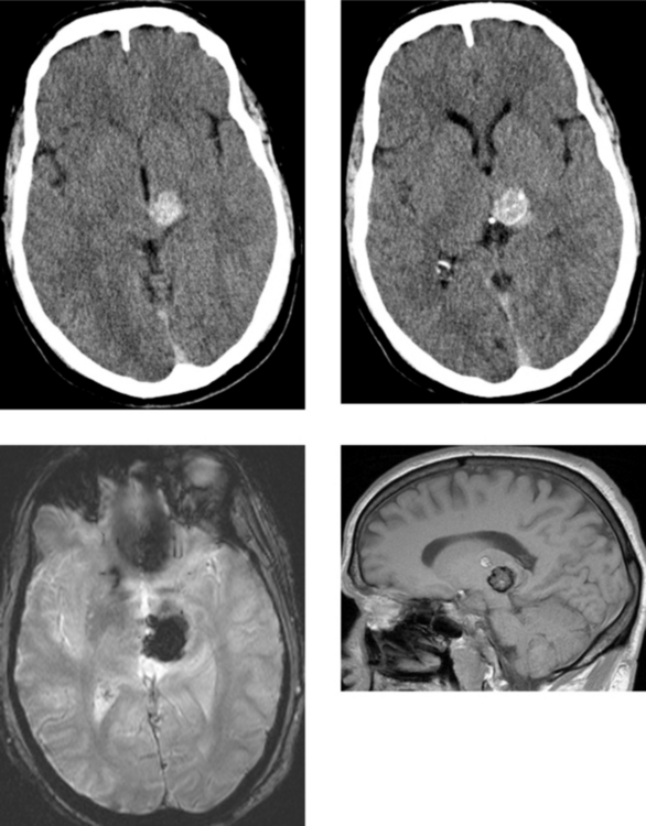
This case shows the typical appearance of an uncomplicated cavernous malformation involving the upper brainstem or cerebral peduncle and left thalamus. In the absence of edema, hemorrhage within the cavernous malformation is unlikely. The differentiation of a cavernous malformation from a hemorrhagic neoplasm on MR imaging occasionally can be difficult in the face of acute hemorrhage, particularly in the presence of edema and mass effect. Several imaging features may help to distinguish these two lesions. Findings favoring a cavernous malformation include focal heterogeneous high signal intensity, representing methemoglobin; a complete hypointense peripheral ring, representing hemosiderin, as in this case; and the absence of enhancing solid tissue. When all else fails, follow-up imaging in 4 to 6 weeks may be performed to assess for the expected temporal evolution of hemorrhage when secondary to a cavernous malformation.


Cavernous malformations may be present in up to 5% of the population. Most are located superficially in the cerebrum and are often closely associated with the adjacent subarachnoid space. They may occur deep within the cerebral hemispheres, although this is less common. Cavernous malformations occur less frequently in the infratentorial compartment. The most common brainstem location is the pons. Many are incidental asymptomatic lesions detected on imaging performed for other indications, as in this patient with traumatic injury. When lesions are symptomatic, symptoms may be related to lesion location or acute hemorrhage. In the cerebrum, the most common presentation is seizures. In the infratentorial compartment, neurologic deficits may occur on the basis of acute hemorrhage, thrombosis, or progressive enlargement of a cavernous malformation related to recurrent hemorrhages.


CASE 163 Capillary Telangiectasia and Developmental Venous Anomaly of the Brainstem


Comment


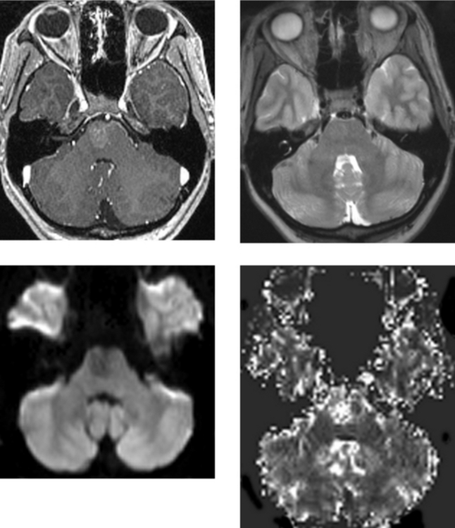
Capillary telangiectasias represent a cluster of abnormally dilated capillaries with intervening normal brain tissue. They usually represent clinically silent lesions that are detected on imaging studies acquired for unrelated reasons. Angiographically, they are most often occult. This case nicely illustrates the typical appearance of a capillary telangiectasia on MR imaging: a poorly demarcated region of “feathery” contrast enhancement, corresponding T2W images that show no significant associated signal abnormality, and gradient echo susceptibility images that show hypointensity in the lesion. It has been postulated that T2W shortening on gradient echo imaging may be related to intravascular deoxyhemoglobin from stagnant blood flow. In this case, regional cerebral blood volume shows increased flow that is believed to reflect pooling of blood in dilated capillaries and venules. Capillary telangiectasias may coexist with other vascular malformations, including cavernomas and DVAs.


DVAs are typically incidental vascular malformations representing an aberration in venous drainage. Within the venous network is intervening normal brain tissue, and no arterial elements are associated with these lesions. The angiomas are composed of a tuft of enlarged venous channels that drain into a common venous trunk, which then subsequently drains into the deep or superficial venous system. Typically, the lesions are clinically silent, although they may be associated with intracranial hemorrhage. These lesions have a characteristic MR imaging appearance, representing a cluster of veins oriented in a “radial” pattern that drain into a large central vein. There is usually no significant signal abnormality in the adjacent brain parenchyma. Angiographically, the arterial and capillary phases are normal, and there may be opacification of the DVA during the venous phase.


CASE 164 Central Neurocytoma


Comment


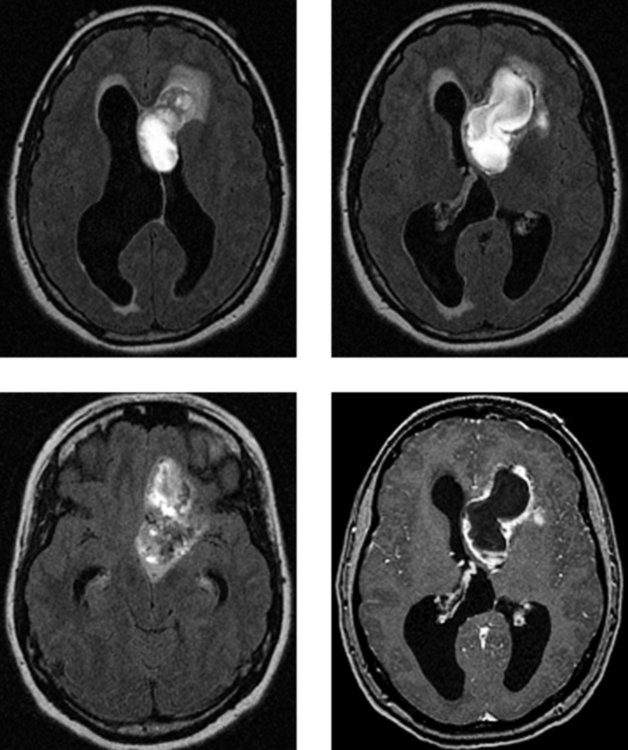
Central neurocytomas typically have a homogeneous cell population with neuronal differentiation. These benign neuroepithelial neoplasms occur in young and middle-aged adults. Patients may be asymptomatic, or may present with headache and signs of increased intracranial pressure, frequently due to hydrocephalus, as in this case. Central neurocytomas arise most commonly within the body of the lateral ventricle (less frequently, the third ventricle), adjacent to the septum pellucidum and foramen of Monroe. They have a characteristic attachment to the superolateral ventricular wall. Most are confined to the ventricles, although occasionally parenchymal extension may occur, as in this case, where there is growth into the frontal lobe. These features may help to distinguish neurocytomas from other intraventricular tumors, such as astrocytoma, giant cell astrocytoma, ependymoma, intraventricular oligodendroglioma, and meningioma. Preoperative diagnosis of central neurocytoma may help in planning therapy, because this tumor has a better prognosis than other intraventricular tumors arising in this area.


On CT and MR imaging, neurocytomas typically are heterogeneous masses that contain multiple cysts. They are well demarcated, with smooth, lobulated margins and moderate vascularity. Most neurocytomas have calcifications. On MR imaging, the more solid component of these tumors tends to follow the signal characteristics of gray matter. Signal voids may be related to calcification or tumor vascularity. Contrast enhancement is variable, ranging from none to moderate. On imaging and conventional pathologic evaluation (light microscopy), these tumors are frequently indistinguishable from oligodendrogliomas. The distinction between these two neoplasms is important because central neurocytomas have a more benign course and treatment may differ. Although neurocytomas have a favorable prognosis, malignant variants and recurrences may rarely occur.


CASE 165 Marchiafava-Bignami Disease


Comment


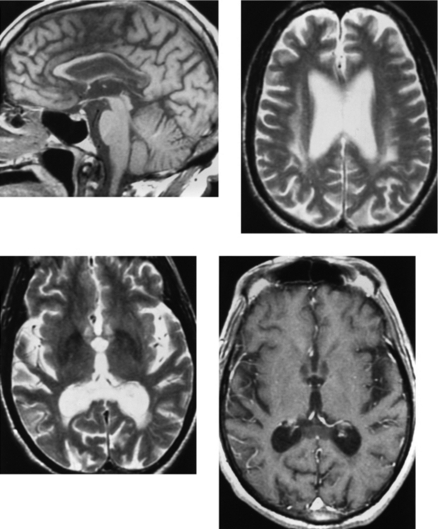
Marchiafava-Bignami disease is a toxic demyelinating disorder initially described by two Italian pathologists. They identified it at autopsy in three patients with chronic alcoholism who presented in status epilepticus that subsequently progressed to coma. All three patients consumed large quantities of red wine. This has also been described in patients with significant nutritional deficiencies, and it has been described in other populations and with other alcoholic beverages. This diagnosis should be considered in patients with acute encephalopathy or progressive dementia and alcoholism. The disease may present acutely, with rapid deterioration, or may exist in a chronic form over a period of years. It is most commonly seen in men, and usually occurs in the third to fifth decade.


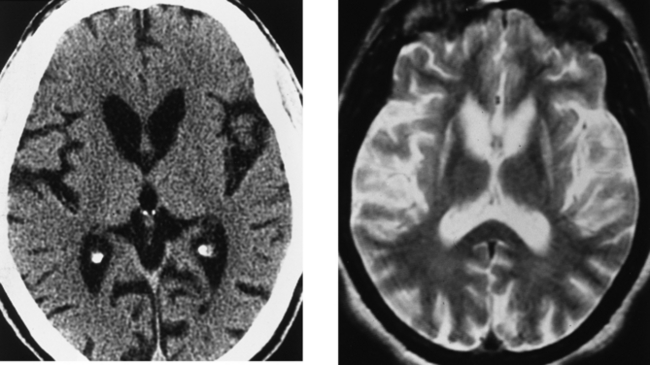
On pathologic evaluation, Marchiafava-Bignami disease is typified by demyelination and necrosis, and it occurs most commonly in the corpus callosum; however, there may be extensive demyelination involving multiple areas of the brain, as in this case, including the deep and periventricular white matter, as well as other commissural fibers. Occasionally, this may involve the subcortical white matter, but typically, the subcortical U-fibers are spared. Sagittal T1W images are valuable in assessing these patients because these images show the extensive callosal atrophy and the associated focal necrosis as demarcated regions of signal abnormality that are hypointense on T1W imaging and hyperintense on T2W imaging. This case nicely demonstrates all of these findings. The acute form of this disease presents with seizures, muscular hypertonia, dysphagia, and coma, and it is often fatal. In the acute form, diffusion-weighted images show hyperintensity (restricted diffusion), and there may be enhancement in affected regions of white matter. In the absence of a patient history, it would be difficult to distinguish this diagnosis from the other demyelinating processes that are more common, such as multiple sclerosis. However, Marchiafava-Bignami disease should be considered in patients with encephalopathy and a history of chronic alcoholism.


CASE 166 Metastatic Carcinoid to the Orbital Extraocular Muscles


Comment


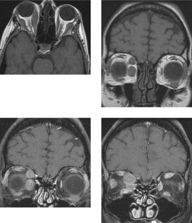
Carcinoid tumors arise from Kulchitsky cells that originate in the neural crest. Most carcinoids arise in the gastrointestinal tract or the lung. This case represents metastatic carcinoid to the orbit, specifically, to the extraocular muscles. With rare exception, the reported metastatic carcinoid tumors to the uvea all developed from primary bronchial carcinoids. In contrast, the vast majority of reported orbital metastases arose from ileal carcinoids. The metastatic potential of carcinoid tumors is related to the site of origin and to tumor size, with histologic features playing a lesser role. In symptomatic patients with intestinal carcinoid, more than 90% have metastatic disease, which is most common to the lymph nodes and liver. Tumors larger than 1 cm are more likely to metastasize.


Metastatic disease to the orbits is common. The imaging diagnosis of orbital metastases as the first manifestation of metastatic disease is not uncommon. For every case of clinically evident orbital metastases, there are several asymptomatic cases that go unrecognized. The most common location for orbital metastases is the globe, usually involving the region of the choroid and retina. Ocular metastases characteristically involve the uveoscleral region. The most common tumors to metastasize to the globe are breast and lung carcinoma in adults. Outside of the globe, orbital metastases most often are extraconal and are often related to bone metastases. Intraconal metastatic disease is usually related to direct extension of an ocular metastasis. Clinical manifestations of ocular metastases are variable. Some patients may be asymptomatic, whereas others may have proptosis, blurred vision, pain, or ophthalmoplegia, depending on the site of involvement.


CASE 167 Joubert’s Syndrome


Jan 22, 2016 | Posted by in NEUROLOGICAL IMAGING | Comments Off on Challenge

Full access? Get Clinical Tree

Get Clinical Tree app for offline access