Chapter 6 Musculoskeletal
Paul J. Spicer
6 Questions and Answers
Refer to the following case for questions 6.1 to 6.3.
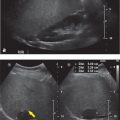
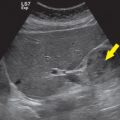
Case 1 A 48-year-old male has Achilles tendon pain. A single long axis (LAX) image of the Achilles tendon is provided. The tendon is noted by the star in the image.

Question 6.1 What is the most appropriate diagnosis for the appearance of this tendon?
Normal tendon.
Tendinosis.
Partial-thickness tear.
Intrasubstance tear.
Full-thickness tear.
Question 6.2 What additional finding is noted on the image, annotated with a vertical arrow, which is often associated with pain?
Retrocalcaneal bursitis.
Subcutaneous calcaneal bursitis.
Calcium hydroxyapatite crystals.
Calcium pyrophosphate crystals.
Monosodium urate crystals.
Question 6.3: The patient requests an Achilles tendon injection for symptom relief and potential expedite healing. Options of corticosteroid injection and platelet-rich plasma (PRP) injection are discussed. Which of the following options best describes the appropriate site of injection?
Corticosteroid—peritendinous, PRP—intratendinous.
Corticosteroid—peritendinous, PRP—peritendinous.
Corticosteroid—intratendinous, PRP—intratendinous.
Corticosteroid—intratendinous, PRP—peritendinous.
Corticosteroid and PRP may be injected either intratendinous or peritendinous.
Answer 6.1:
B. Correct. The single long axis image depicts the Achilles tendon immediately deep to the skin surface, as annotated by the star. The echogenic shadowing structure along the right side of the image is the calcaneus at the tendon insertion. The image demonstrates thickening and hypoechogenicity of the tendon. This appearance is typical of tendinosis. Within the tendon are fibrils, which are the thin hyperechoic lines visualized in length on long axis images. The individual fibrils of the tendon remain intact, without fiber disruption. Therefore, a tear is not visualized.
A. Incorrect. A normal tendon is a thin structure which is hyperechoic. On long axis images, the Achilles tendon is immediately deep to the skin surface. The tendon in this case is much thicker and more hypoechoic than normal, therefore it is not a normal tendon.
C. Incorrect. The fibrils within the tendon remain intact, therefore no tear is present. A partial-thickness tear would result in disruption of a portion of the fibrils on either the skin surface or the pre-Achilles fat pad side of the tendon.
D. Incorrect. The fibrils within the tendon remain intact, therefore no tear is present. An intrasubstance tear would result in disruption of the fibrils within the tendon but without extension to the skin surface side or pre-Achilles fat pad side of the tendon.
E. Incorrect. The fibrils within the tendon remain intact, therefore no tear is present. A full-thickness tear of the Achilles tendon results in a complete tear through the tendon with or without retraction.
Answer 6.2:
A. Correct. There is distention of the retrocalcaneal bursa in the provided image, as annotated bythe vertical arrow. This is noted immediately deep to the tendon and along the superior surface of the calcaneus. It is visualized as a rounded anechoic to hypoechoic structure in the center of the image. This bursa normally resides between the Achilles tendon and the calcaneus; however, when it is distended the bursa extends superior to the calcaneus.
B. Incorrect. Subcutaneous calcaneal bursitis is distention of the bursa located between the skin surface and the Achilles tendon, which is not present in this case.
C, D, E—Incorrect. Crystals are echogenic structures which may be within or outside of a tendon. No crystals are noted in this case.
Answer 6.3:
A. Correct. Corticosteroid injections for tendons, including the Achilles tendon, should be injected peritendinous. An intratendinous injection of corticosteroids weakens the tendon and increases the potential of tendon rupture. Plasma-rich protein (PRP) injections, however, should be performed intratendinous. This is typically performed in conjunction with tenotomy, both of which have a positive effect on tendon healing. The PRP material is able to aid in healing if it is injected within the tendon.
B. Incorrect. PRP should be injected intratendinous, not peritendinous.
C. Incorrect. Corticosteroids should be injected peritendinous, not intratendinous.
D. Incorrect. Corticosteroids should be injected peritendinous, not intratendinous. PRP should be injected intratendinous, not peritendinous.
E. Incorrect. Corticosteroids should be injected peritendinous and PRP should be injected intratendinous, not vice versa.
Refer to the following case for questions 6.4 to 6.6.
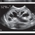
Case 2 A 65-year-old woman has medial epicondyle pain of the elbow. Two consecutive LAX images of the medial epicondyle and common flexor tendons are provided. The flexor tendon is annotated with the downward arrow in both images.

Question 6.4: What is the most appropriate diagnosis for the appearance of the tendon?
Normal tendon.
Tendinosis.
Tendinitis.
Intrasubstance tear.
Full-thickness tear.
Question 6.5: Which of the following changes at the tendon’s cortical bone insertional site is associated with an abnormal tendon?
Increased concavity.
Volume loss.
Flattening.
Increased convexity.
Enthesophyte formation.
Question 6.6: The patient in this case requests percutaneous tenotomy for treatment. How does tenotomy help the healing process of a tendon?
Creates localized bleeding.
Creates neovascularity.
Creates enthesophyte formation.
Prevents fibroblast proliferation.
Prevents collagen formation.
Answer 6.4:
D. Correct. An intrasubstance tear is depicted in the ultrasound as anechoic or hypoechoic focal areas within the tendon. A tear leads to disruption of the echogenic fibrils within the tendon. Intrasubstance tears remain localized within the substance of the tendon while the borders of the tendon on both the deep and superficial surface remain intact. In this case, the tendon which is immediately superficial to the shadowing cortex of the medial epicondyle and is annotated by the downward arrow has focal anechoic tears within the tendon. The deep and superficial surface of the tendon, however, are intact.
A. Incorrect. A normal tendon is hyperechoic with a fiber-like appearance or linear fibrillations within the tendon. This pattern is homogeneous throughout in a normal tendon. In this case, there are focal hypoechoic and anechoic areas within the tendon, suggesting the tendon is abnormal.
B. Incorrect. Tendinosis is depicted at ultrasound imaging as a thickened or swollen, and hypoechoic tendon. The echogenic fibrillations within the tendon remain intact and without a tear.
C. Incorrect. Tendinitis is not a term typically used to describe a tendon, because no acute inflammatory cells are noted within the tendon at biopsy. The term tendinosis is the preferred term.
E. Incorrect. Full-thickness tears are disruptions of the fibrils within the tendon along the superficial surface, the entire intrasubstance portion of the tendon, and the deep surface. The tendon may be retracted.
Answer 6.5:
E. Correct. Enthesophyte formation and irregularity of the cortex of the bone at the insertional site of the tendon often occurs in the setting of an abnormal tendon. This is best seen in image (a). The enthesophytes may intentionally be broken or scrapped during tenotomy to aid in the healing response.
A. Incorrect. Increased cortex concavity of the bone is not associated with tendon abnormalities. Focal pitting or cystic change at the cortex is associated with tendon abnormalities, but the entire cortex does not undergo increased concavity in these cases.
B. Incorrect. Volume loss of the cortex is not associated with tendon abnormalities.
C. Incorrect. Flattening of the cortex of the bone insertional site does not occur, instead enthesophytes and irregularity of the cortex may occur in the setting of an abnormal tendon.
D. Incorrect. Increased cortex convexity of the bone is not associated with tendon abnormalities, instead enthesophytes and irregularity of the cortex may occur in the setting of an abnormal tendon.
Answer 6.6:
A. Correct. Percutaneous tenotomy is also referred to as fenestration or dry needling of a tendon. It helps elicit the healing response by encouraging localized bleeding that leads to fibroblast proliferation and ordered collagen formation. The hypoechoic portion of the tendon, as well as any anechoic cleft, is targeted. If neovascularity is present within the tendon it too is targeted. Tenotomy involves repeatedly passing the needle through the abnormal portions of the tendon to elicit localized bleeding within the tendon.
B. Incorrect. Tenotomy disrupts, not creates, neovascularity.
C. Incorrect. Enthesophytes may also be scrapped from the cortical surface of the bone during tenotomy. Tenotomy does not attempt to create enthesophyte formation.
D. Incorrect. Tenotomy encourages, does not prevents fibroblast proliferation.
E. Incorrect. Tenotomy encourages, does not prevents collagen formation.
Refer to the following case for questions 6.7 to 6.9.
Case 3 A 30-year-old male has the following images of his right supraspinatus tendon in the LAX (a) and short axis (SAX; b). The supraspinatus tendon is labeled by the star on the images.

Question 6.7: What is noted by the vertical arrow on both the SAX and LAX images?
Greater tuberosity.
Lesser tuberosity.
Hyaline cartilage.
Fibrocartilage.
Subacromial-subdeltoid bursa.
Question 6.8: What is noted by the horizontal arrow on the SAX image?
Greater tuberosity.
Lesser tuberosity.
Hyaline cartilage.
Fibrocartilage.
Subacromial-subdeltoid bursa.
Question 6.9: These images were obtained using the modified Crass technique. The purpose of this technique is to bring the tendon out from underneath which structure?
Clavicle.
Coracoid.
Acromion.
Coracohumeral ligament.
Coracoacromial ligament.
Answer 6.7:
A. Correct. The greater tuberosity is the site of insertion of the supraspinatus, infraspinatus, and teres minor tendons. It is devoid of hyaline articular cartilage and the echogenic surface is the cortex of the greater tuberosity. It is labeled with the vertical arrow on both the LAX and SAX images.
B. Incorrect. The lesser tuberosity is the site of insertion of the subscapularis tendon. It is not included in the provided images.
C. Incorrect. The articular surface of the humerus and glenoid is lined by hyaline articular cartilage. The greater tuberosity, however, is not part of the articular surface of the humerus and is, therefore, devoid of hyaline articular cartilage.
D. Incorrect. The fibrocartilage structure within the shoulder joint is the glenoid labrum. It lines the glenoid and helps stabilize the glenohumeral joint. This is not associated with the greater tuberosity.
E. Incorrect. The subacromial-subdeltoid bursa is located on the image between the deltoid muscle and the supraspinatus tendon, labeled with a horizontal arrow on the SAX image. This is not associated with the greater tuberosity.
Answer 6.8:
E. Correct. The subacromial-subdeltoid bursa is located on the images between the deltoid muscle and the supraspinatus tendon, labeled with a horizontal arrow on the SAX image. It is not well visualized in normal individuals, seen as a thin anechoic structure between the deltoid and the tendon; however, in the case of bursitis it may become distended with fluid and is better appreciated. A distended bursa can also lead to subacromial impingement on dynamic maneuvers.
A. Incorrect. The greater tuberosity is the site of insertion of the supraspinatus, infraspinatus, and teres minor tendons. It is devoid of hyaline articular cartilage and the echogenic surface is the cortex of the greater tuberosity. It is labeled with the vertical arrow on both the LAX and SAX images. It is not associated with the subacromial-subdeltoid bursa.
B. Incorrect. The lesser tuberosity is the site of insertion of the subscapularis tendon. It is not included in the provided images.
C. Incorrect. The articular surface of the humerus and glenoid is lined by hyaline articular cartilage. This is not associated with the subacromial-subdeltoid bursa.
D. Incorrect. The fibrocartilage structure within the shoulder joint is the glenoid labrum. It lines the glenoid and helps stabilize the glenohumeral joint. This is not associated with the subacromial-subdeltoid bursa.
Answer 6.9:
C. Correct. The purpose of both the Crass and modified Crass techniques is to bring the supraspinatus and infraspinatus tendons out from under the acromion. The tendons normally reside beneath the acromion; however, the acromion obscures evaluation of the tendons in the resting position. By bringing the tendons out from under the acromion, the tendons can be visualized. This is done by either having the patient put their ipsilateral hand behind the back (Crass technique) or in their back pocket (modified Crass technique). These techniques are sometimes referred to as the Middleton technique.
A. Incorrect. The clavicle does not interfere with visualization of the rotator cuff tendons.
B. Incorrect. The coracoid may obscure a portion of the subscapularis tendon or muscle, but it does not obscure the supraspinatus or infraspinatus tendons.
D. Incorrect. The coracohumeral ligament does not obscure evaluation of the supraspinatus and infraspinatus tendons.
E. Incorrect. The coracoacromial ligament does not obscure evaluation of the supraspinatus and infraspinatus tendons. This ligament does reside superficial to the musculotendinous junction of the supraspinatus and infraspinatus tendons; however, the ultrasound beam is able to penetrate the ligament and therefore it does not obscure the underlying musculotendinous junctions.
Refer to the following case for questions 6.10 to 6.12.
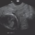
Case 4 A 55-year-old male has shoulder pain. LAX and SAX images of the supraspinatus tendon are provided.

Question 6.10: What is denoted by the structure labeled with the star on the above LAX and SAX images.
Supraspinatus tendon.
Infraspinatus tendon.
Subscapularis tendon.
Deltoid muscle.
Biceps muscle.
Question 6.11: What is the most appropriate diagnosis for the appearance of the supraspinatus tendon?
Normal tendon.
Tendinosis.
Partial-thickness articular-sided tear.
Partial-thickness bursal-sided tear.
Full-thickness tear.
Question 6.12: A 45-year-old patient presents with anterior shoulder pain. Which tendon is most likely affected?
Supraspinatus.
Infraspinatus.
Teres minor.
Short head of the biceps.
Long head of the biceps.
Answer 6.10:
D. Correct. In the provided images, the supraspinatus and infraspinatus tendons are not visualized due to full-thickness, retracted tears. The star indicates the deltoid muscle which is occupying the space adjacent to the hyaline articular cartilage and the cortex of the humeral headwhere the supraspinatus and infraspinatus tendons would normally reside if intact.
A. Incorrect. The supraspinatus tendon is torn and retracted. The stump of the tendon is not included in the provided images.
B. Incorrect. The infraspinatus tendon is torn and retracted. The stump of the tendon is not included in the provided images.
C. Incorrect. The subscapularis is not included in the provided images. The subscapularis tendon is best visualized with the lesser tuberosity externally rotated, however in this case, the greater tuberosity is shown on the provided images.
D. Incorrect. The biceps muscle is visualized overlying the humerus within the upper arm. The provided images are of the shoulder at the greater tuberosity.
Answer 6.11:
E. Correct. The supraspinatus tendon is not visualized on the provided images resulting from a full-thickness, retracted tendon tear. The stump of the tendon is not included on the images. Full-thickness tears result from a tear that extends from the bursal surface to the articular surface of the tendon. If the tear is also completely through the tendon from anterior to posterior, as noted on the SAX image, the stump may retract and therefore may not be visible on the images.
A. Incorrect. A normal tendon is hyperechoic. In the provided images, no supraspinatus tendon is visualized due to a full-thickness, retracted tear.
B. Incorrect. In tendinosis, the tendon swells or thickens, and becomes hypoechoic relative to a normal tendon. The tendon is intact. However, in the provided images, no supraspinatus tendon is visualized and therefore this is not compatible with tendinosis.
C, D— Incorrect. In partial-thickness tears, the tendon remains visible with a tear along the articular or bursal surface or within the substance of the tendon. No supraspinatus tendon is visualized in the provided images, therefore, this is not a partial-thickness tear.
Answer 6.12:
E. Correct. The long head of the biceps tendon is located in the anterior shoulder and is a common cause of pain leading to ultrasound examination of the shoulder. Pain may be the result of tendinosis, rupture, subluxation, or dislocation. The subscapularis tendon is the other anterior shoulder tendon and it too may be a source of pain.
A. Incorrect. The supraspinatus tendon and subacromial-subdeltoid bursa are part of the anterolateral shoulder ultrasound, and therefore are not associated with anterior shoulder pain.
B, C—Incorrect. The infraspinatus tendon and teres minor tendon are part of the posterior shoulder ultrasound examination, and therefore are not associated with anterior shoulder pain.
D. Incorrect. Examination of the short head of the biceps tendon is not part of the routine shoulder ultrasound. This tendon is also a less common cause of pain relative to the long head of the biceps tendon.
Refer to the following case for questions 6.13 to 6.15.
Case 5 A 45-year-old male has posterior elbow swelling and pain. Two images of the site of pain are provided.

Question 6.13: Based on the provided images, what is the most likely diagnosis?
Cellulitis.
Myositis.
Bursitis.
Fasciitis.
Osteomyelitis.
Question 6.14: Which crystal disease would most likely be responsible for the imaging findings?
Calcium pyrophosphate.
Hydroxyapatite.
Amyloidosis.
Uric acid.
Oxalosis.
Question 6.15 Which elbow joint recess is the most sensitive location for identification of joint fluid?
Anterior coronoid fossa recess.
Anterior radial fossa recess.
Posterior olecranon fossa recess.
Annular joint recess.
Sacciform recess.
Answer 6.13:
C. Correct. The complex fluid-filled structure with surrounding hyperemia overlying the olecranon is the olecranon bursa. The olecranon is the shadowing structure within the middle of each image. Bursal distention of the olecranon bursa may be the result of trauma, infection, rheumatoid arthritis, or gout. In this example, the cause was infection of the bursa.
A. Incorrect. The skin and subcutaneous tissues overlying the olecranon bursae are not well-visualized due to the positioning of the focal zones. There is a subtle component of cellulitis, however this is not the predominant finding in this case.
B. Incorrect. Muscle is not clearly visible in this example. There is no evidence of myositis in this case.
D. Incorrect. Fascia is not clearly visible in this example. There is no evidence of fasciitis in this case.
E. Incorrect. The cortex of the olecranon is intact. There is no evidence of osteomyelitis in this example.
Answer 6.14:
D. Correct. Olecranon bursitis may occur as a result of uric acid deposition within the bursa in the setting of gout. The tophi within the bursa will be hyperechoic, which is not seen in this case since this example is the result of infection. Hyperemia and adjacent cortical erosions of the olecranon may additionally be noted.
A. Incorrect. Calcium pyrophosphate, also referred to as calcium pyrophosphate deposition disease (CPPD), does not typically involve the olecranon bursa. Instead, it typically involves the hyaline or fibrocartilage of joints such as the knee and wrist.
B. Incorrect. Hydroxyapatite, which often causes calcific tendinosis, does not typically involve the olecranon bursa.
C. Incorrect. Amyloidosis is not a crystal disease. Instead, it is deposition of a fibrous protein that can lead to amyloid arthropathy. It does not typically involve the olecranon bursa.
E. Incorrect. Oxalosis is the result of calcium oxalate supersaturation in the urine, which can in turn affect osseous structures. However, it does not typically involve the olecranon bursa.
Answer 6.15:
C. Correct. The most sensitive location to evaluate for joint fluid, or an effusion, is the posterior olecranon recess. When imaged in the sagittal plane, superior and posterior displacement of the hyperechoic fat pad will be noted.
A, B—Incorrect. The anterior recesses may distend with fluid in the setting of an effusion; however, these are not the most sensitive location.
D. Incorrect. The annular recess may distend with fluid in the setting of an effusion; however, this is not the most sensitive location.
E. Incorrect. The sacciform recess is an extension of the capsule of the elbow joint at the neck of the radius. This is not the most sensitive site for evaluation of joint fluid within the elbow.
Refer to the following case for questions 6.16 to 6.18.
Case 6 A 45-year-old female has a palpable finding on the palmar surface of her hand near the third digit metacarpophalangeal joint. SAX (a) and LAX images (b) are provided below.

Question 6.16: Based on the provided images, what is the most likely diagnosis?
Giant cell tumor of the tendon sheath.
Dupuytren’s contracture.
Tenosynovitis.
Ganglion.
Foreign body granuloma.
Question 6.17: What does this diagnosis represent?
It is thickening of the palmar aponeurosis.
It is a soft-tissue reaction surrounding a foreign body.
It is a localized form of pigmented villonodular tenosynovitis.
It is synovial thickening within the tendon sheath.
It is a cyst associated with trauma, degeneration, or idiopathic origin.
Question 6.18: Synovial hypertrophy and complex fluid within a joint may appear similar. Which of the following additional findings suggests a diagnosis of synovial hypertrophy instead of complex fluid within the joint?
Joint recess compressibility.
Increased blood flow on power Doppler.
Redistribution of joint contents with transducer pressure.
Hypoechoic appearance.
Focally located within one joint recess.
Answer 6.16:
B. Correct. Dupuytren’s contracture is the result of thickening of the palmar aponeurosis. The mass in the images, noted by the horizontal arrows, is in continuity with the palmar aponeurosis. This continuity is best seen on the SAX image as a thin, hypoechoic structure emanating from the mass. On ultrasound it will appear as an elongated, hypoechoic mass superficial to a flexor tendon sheath and without flow on color or power Doppler.
A. Incorrect. Giant cell tumor of the tendon sheath is a localized form of pigmented villonodular tenosynovitis. The mass is in contact with the tendon sheath, however the tendon is able to glide within the tendon sheath independent of the mass. This mass will have internal flow on color or power Doppler.
C. Incorrect. Tenosynovitis is abnormal fluid distention or synovial thickening within a tendon sheath. This may have increased vascularity on color or power Doppler.
D. Incorrect. A ganglion is a cystic structure. Ganglion are the most common masses of the hand and wrist and are benign. The most common location is along the dorsum of the wrist, adjacent to the scapholunate ligament.
E. Incorrect. A foreign body granuloma would have a foreign body within it, unless it has recently been removed. There is no echogenic foreign body within this mass. Also, foreign body granulomas will have increased vascularity on color or power Doppler.
Answer 6.17:
A. Correct. Thickening of the palmar aponeurosis is a description of Dupuytren’s contracture.
B. Incorrect. Soft-tissue reaction surrounding a foreign body is the description of a foreign body granuloma.
C. Incorrect. A localized form of pigmented villonodular tenosynovitis is the description of a giant cell tumor of the tendon sheath.
D. Incorrect. Synovial thickening within the tendon sheath is the description of tenosynovitis.
E. Incorrect. A cyst associated with trauma, degeneration, or idiopathic origin is the description of a ganglion.
Answer 6.18:
B. Correct. Increased blood flow on power Doppler can be seen with synovial hypertrophy but is not usually seen within complex fluid.
A. Incorrect. Joint recess compressibility is typical of complex fluid. When compressed with the transducer, the fluid will compress away from the transducer, however synovial hypertrophy will not.
C. Incorrect. Redistribution of joint contents with transducer pressure is typical of complex fluid but is not seen in the setting of synovial hypertrophy. Similar to join recess compressibility, if pressure is applied with the transducer, the fluid can redistribute throughout the joint.
D. Incorrect. Both synovial hypertrophy and complex fluid may have a hypoechoic appearance.
E. Incorrect. Complex fluid may be focally located within one joint recess, however synovial hypertrophy is usually present throughout the joint.
Refer to the following case for questions 6.19 to 6.21.
Case 7 A 30-year-old female has ankle pain and a palpable mass. LAX (a) and SAX color Doppler images (b) are provided.

Question 6.19: Based on the images what is the most likely diagnosis?
Ganglion.
Lipoma.
Lymph node.
Soft-tissue sarcoma.
Nerve sheath tumor.
Question 6.20: What do the vertical arrows indicate?
Fat.
Muscle.
Vessel.
Nerve.
Fascia.
Question 6.21: What is the most common soft-tissue tumor?
Angiolipoma.
Lipoma.
Lipoblastoma.
Liposarcoma.
Hibernoma.
Answer 6.19:
E. Correct. A nerve sheath tumor is a solid soft-tissue mass that is in continuity with a peripheral nerve. These masses are typically hypoechoic and may have low-level internal echoes. They are round or oval and are well defined. The mass often will have increased through transmission, as this example does, which may cause it to be mistaken for a cyst. However, color or power Doppler imaging will demonstrate internal vascularity which will distinguish it from a cyst.
A. Incorrect. A ganglion is a cystic mass, however they will not have internal vascularity as the mass in this case does.
B. Incorrect. A lipoma may be present within the subcutaneous tissues or within a muscle. The echogenicity may vary depending on the location of the mass, either within the subcutaneous tissues or within a muscle, and the amount of fibrous tissue within the mass. Lipomas within a muscle or those which have more fibrous tissue will appear hyperechoic. The example in this case, however, is more hypoechoic and more round than is typical of a lipoma.
C. Incorrect. Lymph nodes are typically reniform in shape with a fatty echogenic hilum. Blood flow to the lymph node is usually through the hilum. The cortex is hypoechoic. The example in this case is not compatible with a normal lymph node.
D. Incorrect. Soft-tissue sarcomas are typically hypoechoic. They may have areas which are anechoic due to necrosis within the mass. They have increased vascularity on color or power Doppler imaging and have increased posterior through transmission. They, however, are not in continuity with a nerve.
Answer 6.20:
D. Correct. The vertical arrows point to the nerve as it enters and exits the mass. This is characteristic of a nerve sheath tumor.
A. Incorrect. The subcutaneous fat within the images is superficial to the mass.
B. Incorrect. No muscle is noted within the images.
C. Incorrect. Small vessels are noted within the color Doppler image; however, these are not depicted by the vertical arrows.
E. Incorrect. No fascia is noted within the images.
Answer 6.21:
B. Correct. Lipomas are the most common soft-tissue tumors, accounting for approximately 50% of allsoft-tissue tumors. They occur most commonly in the fifth through seventh decades of life. They are composed of mature adipose tissue and they resemble subcutaneous fat. They may have a few thin septa, less than 2 mm in thickness.
A. Incorrect. Angiolipoma is a benign neoplasm which presents in the second or third decade of life as multiple, small, tender, subcutaneous nodules within the forearm, upper arm, or trunk.
C. Incorrect. Lipoblastoma is a rare neoplasm of childhood, typically occurring in children less than 3 years of age. They most commonly occur in the extremities as a painless, enlarging mass.
D. Incorrect. Liposarcomas account for approximately 17% of all soft-tissue sarcomas. They usually present in the sixth decade of life as a painless, enlarging mass.
E. Incorrect. Hibernoma is an uncommon benign tumor arising from brown fat. It presents as a painless, slow-growing mass in adults.
Refer to the following case for questions 6.22 to 6.24.
Case 8 A 55-year-old male undergoes an ultrasound-guided joint injection. A single image is provided.

Question 6.22: What artifact is present in the image?
Beam width.
Reverberation.
Mirror image.
Refraction.
Increased through transmission.
Question 6.23: What would be the best way to improve this artifact?
Change the angle of insonation.
Adjust the focal zones.
Use color Doppler.
Increase the depth.
Increase the gain.
Question 6.24: What artifact is created when the ultrasound beam is angled as little as 5 degrees off perpendicular to the target?
Anisotropy.
Shadowing.
Ring-down.
Mirror image.
Beam width.
Answer 6.22:
B. Correct. Reverberation artifact occurs when two parallel highly reflective surfaces are present. The echoes may be reflected back and forth between the two surfaces before ultimately returning to the transducer. Since it takes longer for these echoes to return to the transducer, the ultrasound processor infers that they originated from a deeper location and places them accordingly deeper in the image. In this case, the two highly reflective parallel surfaces are the needle and the cortex of the bone.
A. Incorrect. The width of the ultrasound beam is initially the width of the transducer. However, as it nears the focal zones, the beam narrows and once it is distal to the focal zones it again widens, even wider than the width of the transducer itself. Beam width artifact occurs when the width of the beam is wider than the transducer. Echoes which originate from the portion of the beam that is beyond the width of the transducer are assumed to occur within the narrower transducer width of the beam and are placed within this portion of the image. This is why a large cyst may appear to have debris within the margins of the cyst if that portion of the cyst is outside the normal width of the beam.
C. Incorrect. In mirror image artifact, the primary beam encounters a highly reflective interface. The echo is reflected toward the transducer, however along its path it encounters the backside of a highly reflective surface causing the reflected echo to be reflected again toward the original highly reflected interface, away from the transducer. The original reflective interface again reflects the echo toward the transducer. This results in an image with a duplicated structure which is equidistant from and deep to the strongly reflective interface.
D. Incorrect. Refraction artifact occurs when the ultrasound beam travels through a medium other than human tissue, such as air or fluid. The speed of the ultrasound travels at a different rate in these other media, and therefore the echo is miscalculated and may be placed deeper in the image as a result.
E. Incorrect. Increased through transmission is the result of the ultrasound beam encountering a focal weak attenuating structure in the field. The amplitude of the beam that passed through this weak attenuating structure will be greater than the amplitude of the remainder of the beam in the image at the same depth. This increased amplitude is falsely recorded as increased echogenicity or increased through transmission.
Answer 6.23:
A. Correct. If the angle of insonation of the transducer is changed, or if the angle of the needle is changed, then the needle and cortex of the bone may no longer be parallel. Thus, the reverberation artifact would resolve.
B. Incorrect. Adjusting the focal zones would not resolve the reverberation artifact.
C. Incorrect. The use of color Doppler would not resolve the reverberation artifact.
D. Incorrect. Increasing the depth would not resolve the reverberation artifact.
E. Incorrect. Increasing the gain would not resolve the reverberation artifact.
Answer 6.24:
A. Correct. Anisotropy may occur when the ultrasound beam is angled relative to the target structure. This can occur when the beam is angled as little as 5 degrees. The result is normal tendons may appear hypoechoic due to the artifact, thus mimicking tendinosis. To determine if the hypoechoic appearance of a tendon is real or due to anisotropy artifact, the transducer angle can be altered by rocking the probe back and forth to see if the hypoechoic appearance of the tendon persists. If it does persist it is tendinosis, if not then it is the result of anisotropy artifact. A similar result of this artifact can occur with ligaments and muscle.
B. Incorrect. Shadowing occurs when the ultrasound beam is reflected. This results in an image with an anechoic region deep to the involved interface. This may also occur if the beam is absorbed or refracted. In musculoskeletal ultrasound, this may be encountered with bone, calcification, foreign bodies, or gas.
C. Incorrect. Ring-down artifact is a type of reverberation artifact in which the reflective echoes are more continuous deep to the source, usually the result of gas bubbles. The reflective echoes are tightly associated with each other and they produce an appearance of a continuous, echogenic stripe deep to the source.
D. Incorrect. In mirror image artifact, the primary beam encounters a highly reflective interface. The echo is reflected toward the transducer; however, along its path it encounters the backside of a highly reflective surface causing the reflected echo to be reflected again toward the original highly reflected interface, away from the transducer. The original reflective interface again reflects the echo toward the transducer. This results in an image with a duplicated structure which is equidistant from and deep to the strongly reflective interface.
E. Incorrect. Beam width artifact occurs when the width of the beam is wider than the target. Echoes originating from the portion of the beam that is beyond the width of the transducer are assumed to occur within the narrower transducer width of the beam and places them within this portion of the image. This is why a large cyst may appear to have debris within the margins of the cyst if that portion of the cyst is outside the normal width of the beam. Anisotropy artifact occurs when the ultrasound beam is angled as little as 5 degrees relative to the target structure. It may falsely make a normal tendon appear to have tendinosis. It may also make a normal ligament appear injured.
Related posts:

Stay updated, free articles. Join our Telegram channel

Full access? Get Clinical Tree


