Chest CT
After having discussed normal anatomy of caudal cervical sections (p. 67), normal thoracic anatomy is presented. From this page on, you will find the number codes for the drawings in the rear foldout.
Selection of Image Plane
As a rule, the sections of the thorax are chosen in the transverse or axial plane at thicknesses and steps of 4 to 8 mm. Sections 7 mm thick will overlap by 1 mm, for example, when the patient table is advanced in 5 mm steps. A small topogram ( Fig. 75.1 ) accompanying each sheet of images shows the position of the sections relative to the major anatomic structures of the region. In order not to miss any pathologic changes within the lung (review p. 13), it has become accepted practice to make a hard copy of both soft-tissue and lung windows or to provide a CD with the image data. Each image can therefore be viewed at two different window settings. Again the large number of images necessitates a systematic technique for evaluation so as not to waste time looking randomly back and forth between them.
Systematic Sequence for Readings
The beginner often forgets to check the soft tissues of the thoracic wall because the examination of the mediastinum and the lungs is automatically considered more important. These tissues should therefore be evaluated first. Common sites of abnormality are the breasts and fat in the axilla (2). After this–also using soft-tissue windows–the mediastinum is checked for pathologic masses. The easiest approach is to orient yourself relative to the arch of the aorta (89b), which can be recognized even by the inexperienced ( Fig. 77.3 ). From this point cranially, the major branches are identified to exclude pathologic masses in the upper mediastinum next to the brachiocephalic trunk (88), the left common carotid artery (85), the subclavian artery (87), as well as the brachiocephalic veins (91), superior vena cava (92), trachea (81), or more dorsally, the esophagus (82).
Caudally, the most common sites for enlarged LNs are: at the aortopulmonary window, directly below the bifurcation of the trachea (81a), in the perihilar tissue, posterior to the crura of the diaphragm (=retrocrural) next to the descending aorta (89c). The presence of a few LNs smaller than 1.5 cm in diameter in the aortopulmonary window may be considered normal [19, 41]. Anterior to the aortic arch (89b) LNs of normal size are rarely seen in the CT. The evaluation of the soft-tissue window is complete when the heart (any coronary sclerosis, dilations?) and the lung hila (vessels well defined and not lobulated or enlarged) have been checked. Only now should the radiologist turn to the lung or pleural window.
Since the pleural window is very wide, the marrow of the spinal column as well as the parenchyma of the lung can be examined. It is therefore possible to evaluate bone structure in addition to the pulmonary vasculature. When examining the lung vessels, look for a gradual reduction in their diameter as you proceed from the hilum to the periphery. Pulmonary oligemia is normal only along the margins of the lobes and in the periphery. It is essential to differentiate between cross-sectioned vessels and solid masses by comparing adjacent levels (cf. p. 15). More or less spherical solid masses may indicate intrapulmonary metastases. The checklist will help you read thoracic CTs systematically.
The simultaneous presentation of two window settings in one hard copy (both the lung and the soft-tissue window) has not proved practical because pathologic abnormalities which have density levels between the two would be overlooked. Consult the lung chapter on pages 84ff. for scans in the lung window.
Checklist for Thorax Readings
On the soft-tissue window:
soft tissues, especially:
axillary LNs
breast (malignant lesions?)
mediastinum in four regions:
from the aortic arch cranially (LNs?, thymoma / struma?)
hilar region (configuration and size of vessels, lobulated and enlarged?)
heart and coronary arteries (sclerosis?)
four typical sites of predilection for LNs:
anterior to aortic arch (normal: almost none or < 6 mm)
in the aortopulmonary window (normal: < 4 LNs < 15 mm)
subcarinal (normal: < 10 mm; DD: esophagus)
next to descending aorta (normal: < 10 mm; DD: azygos)
On the lung window:
Parenchyma of the lung:
normal branching pattern and caliber of vessels?
vascular oligemia only at interlobar fissures? bullae?
any suspicious lung foci? inflammatory infiltrates?
Pleura: – plaques, calcification, pleural fluid, pneumothorax?
Bones (vertebrae, scapula, ribs):
normal structure of marrow?
degenerative osteophytes?
focal lytic or sclerotic processes?
stenoses of the spinal canal?
The parenchyma of the thyroid gland (83) should appear homogeneous and clearly defined from the surrounding fat (2). Asymmetry in the diameter of the jugular vein (86) is seen quite often and has no pathologic significance. Orthogonally sectioned branches of the axillary (93) and lateral thoracic (95) vessels must be distinguished from axillary LNs.
If the arms are elevated, the supraspinatus muscle (19) lies medial to the spine of the scapula (53b) and the infraspinatus muscle (20). Usually the pectoralis major (26a) and minor (26b) muscles are separated by a thin layer of fat.


Artifacts (3) will be observed at the level of the thoracic inlet if CM is present in the subclavian vein (87) at the time of data acquisition (cf. Fig. 23.3).
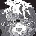
Thoracic CTs are also viewed from caudally. The left lung (84) appears on the right side of the image and vice versa. Beginning at the aortic arch (89b in Figs. 77.2/3 ), the layout of the aortic arch vessels should be thoroughly familiar to you. At the section in Figure 76.1 , the left subclavian artery (87) is seen most posteriorly and can be followed in cranial direction in the images on page 75. In front of the subclavian artery lie the left common carotid artery (85) and the brachiocephalic trunk (88). More to the right and anteriorly are the brachiocephalic veins (91), which form the superior vena cava (92) at the levels of Figures 76.3 to 77.1 . In the fat of the axilla (2), normal LNs (6) are often recognizable by their typical indented shape: the hilum contains fat. At a different angle, the hypodense hilum will appear in the center of an oval. Healthy LNs are well defined and should not exceed 1 cm in diameter in this location ( Figs. 76.1 and 76.3 ).


The azygos vein (104) lies dorsal to the trachea (81) next to the esophagus (82). Directly above the right main bronchus, it arches anteriorly into the superior vena cava (92). Be sure not to confuse the paravertebral azygos vein (104), the hemiazygos vein (105) or accessory hemiazygos (105a) with paraaortic LNs ( Fig. 77.3 ).


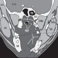
Immediately caudal to the arch of the aorta (89b) is situated the pulmonary trunk (90), which divides into the right (90a) and left (90b) pulmonary arteries ( Fig. 78.2 ). At the level of Figure 78.1 there is the aortopulmonary window, a site of predilection for mediastinal LNs (6). Also check for enlarged LNs or malignant masses in the subcarinal position between the two main bronchi (81b) close to the pulmonary vessels (96) ( Fig. 78.3 ). Near the internal thoracic (mammary) vessels (94) lies the regional lymphatic drainage of the medial parts of the breasts, whereas the lymphatic drainage of the lateral portions of the breasts is primarily to the axillary nodes.


The glandular tissue (72) in the fat of the breasts of the anterior thoracic wall is easily differentiated from skin tumors because of the symmetry ( Figs. 79.1–3 ). The main coronary arteries (77) are also distinguishable in the epicardial fat ( Figs. 79.2/3 ). Develop a clear mental picture of the positions of the azygos vein (104) and the esophagus (82) next to the descending aorta (89c) so that you will later be able to recognize any pathologic LNs close to these structures.


The left atrium (74c) is the most posterior chamber of the heart, whereas the outlet of the left ventricle (74d) and the ascending aorta (89a) lie in the center of the heart. The right atrium (74a) lies on the right lateral side and the right ventricle (74b) anteriorly behind the sternum (56). Only the larger central branches of the pulmonary vessels (96) can be seen on the soft-tissue window. The smaller, more peripheral lung vessels are better judged on the lung window (not shown here).
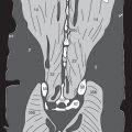
Note the junction between the hemiazygos vein (105) and the azygos vein (104), which must not be confused with a paravertebral lymphoma ( Fig. 80.3 ).


The sections on previous and this page show the opening of the coronary sinus (76) into the right atrium (74a) and sequential sections of the coronary arteries (77). The hypodense epicardial fat (79) must not be mistaken for fluid within the pericardial space. The internal thoracic artery, also known as the internal mammary artery (94), is more and more frequently used in bypass operations. It is surgically anastomosed with the anterior descending branch of the left coronary artery.


The inferior vena cava (80) is seen more caudally ( Figs. 82.1–3 ), and finally the diaphragm (30) appears together with the upper parts of the liver (122). Many radiologists who suspect the presence of a bronchial carcinoma (BC) obtain images to the caudal edge of the liver (see p. 83) because a BC often metastasizes to the liver and the adrenal glands. The caliber of lung vessels near the periphery of the diaphragm is so small that they are not visible on the soft-tissue window, as in the present images. The pattern of the pulmonary vasculature should therefore be examined on the lung windows, which include the negative density values of the Hounsfield scale. Only after this step has been carried out is the evaluation of a chest CT complete.


Test Yourself! Exercise 19:
Write down a concise but complete sequence of all criteria for interpreting a thoracic CT. Then compare your notes with the checklist on page 74 and repeat this exercise from time to time until you remember every criterion.
Related posts:


Stay updated, free articles. Join our Telegram channel

Full access? Get Clinical Tree


