Chest Wall


Chest Wall


Dawn R. Engelkemier

Peter G. Kruk

John Naheedy

Yeun-Chung Chang

Pilar Dies-Suarez

Edward Y. Lee


INTRODUCTION

Pediatric chest wall lesions are common. Abnormalities may arise from any component of the chest wall and from a vast array of conditions including congenital and developmental anomalies, infectious disorders, neoplastic disorders, traumatic lesions, and vascular anomalies. Although there is some overlap with adult pathology, many of these entities are unique to the pediatric population. Imaging plays a critical role in detection, characterization, and management of pediatric chest wall lesions (Table 12.1). This chapter reviews imaging techniques for evaluating the pediatric chest wall and briefly discusses normal anatomy and variants. The remainder of the chapter classifies and describes a spectrum of pediatric chest wall lesions with attention to clinical features, imaging characteristics, and treatment options.


IMAGING TECHNIQUES

Various imaging modalities are currently used for evaluation of chest wall lesions in pediatric patients. The imaging techniques as well as unique advantages and disadvantages of these imaging modalities are discussed in this section and summarized in Table 12.2.


Radiography

After clinical evaluation has been performed, chest radiographs are most commonly the first imaging modality employed to evaluate chest wall lesions in pediatric patients. Radiography is a rapid, widely available, low-cost technique that is relatively easy to acquire.1 Evaluation typically consists of a two-view chest radiograph. In infants, an anterior-posterior (AP) projection is obtained with the patient recumbent, and a cross-table lateral technique is used for the lateral view. Once the child is able to sit with minimal support (around 1 year of age), standard posterior-anterior (PA) and lateral views can be obtained. A grid is used for imaging of adolescent, adult, and large patients.

In addition to standard chest radiographs, dedicated oblique views of the ribs may facilitate characterization of chest wall lesions. Placing a radiopaque marker in the area of concern prior to imaging can help correlate imaging findings with patient symptoms (Fig. 12.1). The patient is positioned erect for imaging of ribs projecting above the diaphragm and recumbent for ribs projecting below the diaphragm. Imaging may include 45-degree right anterior oblique projection for left anterior ribs, 45-degree left anterior oblique projection for right anterior ribs, 30- to 45-degree left posterior oblique for imaging the left axially border, and 30- to 45-degree oblique for imaging the right axillary border.

Some pediatric chest wall lesions have characteristic radiographic findings, and radiographs may be diagnostic. In other cases, radiographs provide valuable information, such as whether a lesion is primarily osseous or soft tissue, and can guide further imaging evaluation.


Ultrasound

Ultrasound (US) may follow radiographs for further evaluation and characterization, or it may be used as a primary modality for superficial lesions, particularly those that are visible or palpable. It has many benefits as an imaging modality, particularly in the pediatric population. US can be performed at the bedside and provides dynamic, real-time information. Sedation and intravenous access are typically unnecessary.

Additionally, patients are not exposed to the potentially harmful effects of ionizing radiation.


TABLE 12.1 Practical Evaluation of Pediatric Chest Wall Lesions


image


Adapted from Karmazyn B, Davis MM, Davey MS, et al. Imaging evaluation of chest wall tumors in children. Acad Radiol. 1998;5(9):642-654, Ref.86


TABLE 12.2 Advantages and Disadvantages of Imaging Modalities for Evaluation of Pediatric Chest Wall Lesions


Modality


Advantages


Disadvantages


Radiography


  • Rapid


  • Accessible


  • Low cost


  • Radiation


  • Limited evaluation of soft tissues


Ultrasound


  • No radiation


  • Dynamic


  • Portable


  • Limited field of view and depth visibility


  • Operator dependent


CT


  • Fast


  • Excellent assessment of lung parenchyma and osseous lesions


  • Radiation


  • May require sedation in young children


MRI


  • Excellent soft tissue characterization


  • No radiation


  • Relatively long acquisition time


  • May require sedation


  • Susceptible to artifacts


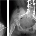
FIGURE 12.1 Oblique rib radiograph with a radiopaque marker placed in area of palpable mass.

The ultrasound technique should be tailored to the child and to the clinical question. Patient positioning is critical for optimizing the acoustic window and maximizing patient comfort, thereby minimizing motion.2,3,4,5 The anatomic area of interest and patient age and size determine transducer selection. Curved or linear array transducers are commonly utilized. Curved transducer probes provide a wide field of view. Linear probes yield superior resolution at shallow depths. High-frequency (7.5 to 15 MHz) transducers are usually appropriate for evaluation of pediatric chest wall lesions as they provide superior spatial resolution but decreased penetration compared to lower frequency transducers. Tissue equivalent standoff material may improve visualization of superficial lesions.

US may identify the location and extent of a lesion including the chest wall layers involved. The cystic, solid, or vascular nature can be evaluated with gray scale and color Doppler imaging. Some benign lesions have a characteristic US appearance that negates the need for further workup.


Computed Tomography

Computed tomography (CT) is a valuable noninvasive diagnostic tool when radiography and/or US is not sufficient, particularly in evaluation of lesions with an osseous component. CT may be necessary for preoperative planning. A simultaneous assessment of the lung parenchyma can be performed.1 Modern multidetector CT (MDCT) provides fast acquisition, high spatial resolution, and excellent image quality. Multiplanar and three-dimensional (3D) reformatted images allow evaluation from many perspectives. In recent years, the need for sedation has decreased in light of the fast scanning times of MDCT. Infants and children younger than 5 years or who are unable to lay still or follow breath holding (BH) instructions may still require sedation.6 Oral chloral hydrate and intravenous pentobarbital are commonly used for moderate (conscious) sedation.7 Thorough presedation evaluation, monitoring, and cardiorespiratory support by trained providers are critical for performing safe sedated imaging.8

In accordance with the “as-low-as-reasonably-achievable” (ALARA) principle, CT in pediatric patients should be performed with the lowest possible radiation dose that maintains diagnostic imaging quality.1 For some indications, such as pectus excavatum or focal rib lesions, limiting the anatomic coverage to the area of concern can decrease radiation exposure. Specific technical parameters depend on the protocol and type of MDCT scanner used; however, in general, tube current and kilovoltage peak should be adjusted based on patient weight.6 MDCT should be performed with a fast table speed, thin detector collimation, and a thin reconstruction interval in order to produce high-quality multiplanar and 3D reconstruction images9 (Table 12.3). Both bone and soft tissue algorithms should be used for reconstruction. Noncontrast examinations are useful to evaluate osseous lesions and identify calcifications. Intravenous contrast is typically used to evaluate soft tissue lesions.1


TABLE 12-3 CT Protocol for Chest Wall Lesions


Anatomic coverage (extent)


From sternal notch to below the diaphragm


Scanner settings


See Table 6.1 in this book


Detector collimation


16 row: ˜0.75 mm 64 row or higher row: ˜0.6 mm


Pitch


1-1.5


Intravenous contrast type


Nonionic: 280-320 mg iodine/mL


Intravenous contrast volume


1-2 mL/kg (up to 100 mL)


Intravenous contrast injection rate


Hand injection: rapid push (˜1 mL/s)


Mechanical (power) injector:


22 gauge: 1.5-2.5 mL/s


20 gauge: 3-4 mL/s


Scan initiation


˜30-40 s after start of contrast injection


Reconstructions (routine viewing)


5 mm × 5 mm


Window width (W) and level (L)


Mediastinum: 400 W and 40 L


Lung: 1,250 W and -500 L


Bone: 1,500 W and 300 L


Reconstructions (3D imaging)


3 mm × 2 mm or 3 mm × 3 mm


Postprocessing techniques


Multiplanar reconstruction and volume-rendered reformation


Magnetic Resonance Imaging

Magnetic resonance imaging (MRI) is complimentary to radiographs, US, and CT when questions remain unanswered. MRI provides excellent soft tissue characterization without radiation exposure and is particularly useful in evaluating potential spinal involvement. Infants and young children (≤5 years) usually require sedation.

The specific technique should be tailored to the patient and clinical question. A multichannel cardiac coil is typically used for infants and children under the age of 5 years. A phased array torso coil is used in older children and adolescents. Respiratory triggering (RT) and breath holding (BH) techniques can decrease artifact from respiratory motion. Prone positioning may be useful for anterior chest wall lesions to decrease respiratory motion in the area of interest.10 Flow compensation and flow presaturation techniques can minimize artifact from vascular motion.

The basic protocol for imaging chest wall masses begins with axial and coronal T2-weighted fat saturation (FS) fast relaxation fast spin-echo sequence with BH or RT followed by axial T1-weighted fast spin-echo sequence. Sagittal inversion recovery MR images may be particularly useful for evaluating paraspinal lesions. Diffusion-weighted sequence help characterize soft tissue lesions. Dynamic 3D MR angiogram
and postcontrast T1-weighted FS sequence provide information about the vascularity of soft tissue masses and vascular anomalies.


Nuclear Medicine

Nuclear medicine studies may occasionally be used for evaluation of pediatric chest wall lesions and as with other imaging modalities should be appropriately tailored. Radiopharmaceutical doses should be kept as low as possible; a balance must be achieved between minimizing radiation and obtaining a quality diagnostic exam in a reasonable amount of time. Longer acquisition times are more susceptible to patient motion. The length of time the child has to lie still can be decreased by separating long dynamic studies into sequential static images to allow the child to move between acquisitions.11

Pediatric administered activity is usually calculated by multiplying the adult reference activity by one of several dose formulas. Body surface area and Webster’s formulas have been commonly used; however, these calculations result in significantly larger administered activities per kilogram for infants and small children who are most susceptible to the effects of ionizing radiation. The North American Consensus Guidelines for Administered Radiopharmaceutical Activities in Children and Adolescents recommend weight-based calculations for most common pediatric nuclear medicine studies in children over 1 year of age. In infants, minimum total doses necessary for an adequate study regardless of weight can be used.12

Bone scintigraphy and fluorodeoxyglucose (FDG) positron emission tomography (PET) may be of particular use in evaluating a variety of pediatric chest wall disorders including infectious, inflammatory, neoplastic, and traumatic lesions and can aid in diagnosis of multifocal disease. More recently, 18F-NaF PET has been used for detection of new bone formation in a variety of skeletal disorders including identification of skeletal metastasis and skeletal injuries in child abuse.13,14


FIGURE 12.2 Skeletal chest wall anatomy.


NORMAL ANATOMY AND VARIANTS

The chest wall extends from the skin to the parietal pleural. It provides vital functions including protection of underlying viscera, support for respiratory function, and framework for the shoulders and arms. The normal chest wall is symmetric and broadens cranially to caudally. The skeleton of the chest wall is composed of the sternum anteriorly and the spinal column posteriorly with twelve thoracic vertebrae and paired ribs (Fig. 12.2). The first seven pairs of ribs extend from the costovertebral articulations posteriorly to the sternocostal articulations anteriorly. The eighth through tenth ribs attach to each other anteriorly by costal cartilage; the eleventh and twelve ribs float anteriorly unattached. The internal mammary, posterior intercostal, lateral thoracic, thoracoacromial, and transverse cervical arteries provide the main blood supply to the chest wall.15

The configuration of the chest wall changes with age. In infancy, the ribs are oriented horizontally. When an upright position is adopted, the ribs begin to slope downward. The adult rib configuration is reached by ˜10 years of age.
Ossification of the skeletal elements begins in utero and continues until the 25th year of life. Due to differences in muscle mass and ossification, the chest wall of infants and children is more elastic and compliant than in adults. This results in lower resting lung volumes and a less efficient respiratory mechanism that predisposes infants and young children to atelectasis. The osseous and soft tissue components gradually become stiffer with age.16


FIGURE 12.3 Layers of the chest wall.

The chest wall can be divided into three layers: a superficial layer of skin and subcutaneous fat; an intermediate layer containing the shoulder girdle and pectoralis muscles; and a deep layer including the sternum, ribs, intercostal space, spine, fascia, and parietal pleural17 (Figs. 12.3 and 12.4). Lesions may arise from any of these layers and their components including the vessels and nerves that course through them.


FIGURE 12.4 Chest wall layers as seen on axial CT image.


SPECTRUM OF CHEST WALL DISORDERS


Congenital and Developmental Anomalies

Congenital and developmental anomalies of the pediatric chest wall are common. Mild chest wall asymmetries occur in approximately one third of children.18 Palpable, but otherwise asymptomatic anterior chest bumps are usually due to anatomic variations; however, they may prompt imaging evaluation for a possible underlying true mass.19 If the lesion is small, nontender, and stable, radiographs likely provide adequate evaluation. In young children, a predominance of nonossified cartilage in the anterior chest wall may make radiographic evaluation difficult; US can be very useful in these cases.


Congenital chest wall anomalies are frequently seen in association with congenital scoliosis. When the scoliosis is due to unilateral vertebral segmentation failure, the chest wall abnormality occurs on the concave side of the scoliosis ipsilateral to the vertebral anomaly.20


Chest Wall Asymmetry


Pectus Excavatum

Pectus excavatum is the most common congenital chest wall deformity with a prevalence of 1.3% to 2.6% in school-age children and a male to female ratio of 5:1.21,22,23 It is characterized by prominent indentation of the lower sternum, which is usually asymmetric to the right, and resulting decrease in AP diameter of the rib cage24 (Table 12.4). The etiology has not been clearly elucidated but may be related to abnormal development of costal cartilages.25 The defect may be noticeable at birth or within the first year, increases in severity during puberty, and stabilizes in adulthood. Associated musculoskeletal abnormalities, particularly scoliosis, are common. Congenital heart disease, osteogenesis imperfecta, muscular dystrophy, and various syndromes including Poland, Pierre Robin, Turner, Ehlers-Danlos, and prune belly syndrome are associated.26 Two-thirds of patients with Marfan syndrome have pectus excavatum; in these cases the deformity tends to present later and is more progressive.27


TABLE 12.4 Key Features of Common Primary Pediatric Chest Wall Lesions


Lesion


Classification


Key Features


Pectus excavatum


Congenital/developmental


  • Lower sternal indentation


  • Decreased anteroposterior diameter rib cage


  • Syndromic associations


Lipoma


Soft tissue benign neoplasm


  • Homogeneous fat density on CT and signal intensity on MRI


Osteochondroma


Osseous benign neoplasm


  • Metaphyseal location


  • Corticomedullary continuity with underlying bone


  • Cartilaginous cap


Ewing sarcoma


Malignant neoplasm


  • Rib location most common


  • Osseous destruction with periosteal reaction and large soft tissue mass


  • Heterogeneous enhancement


Rhabdomyosarcoma


Soft tissue malignant neoplasm


  • Large painful soft tissue mass


  • Osseous erosion


  • Heterogeneous enhancement


Rib fracture


Traumatic


  • May appear more aggressive during healing due to periosteal reaction


  • Suspicious for child abuse without appropriate traumatic history


Venous malformation


Vascular malformation


  • Low flow


  • Soft


  • Compressible


  • Phleboliths


  • Gradual diffuse enhancement


Lymphatic malformation


Vascular malformation


  • Low flow


  • Rubbery


  • Noncompressible


  • Septated cysts with fluid-fluid levels


  • Mild mural enhancement


Hemangioma


Vascular tumor


  • High-flow vascular channels


  • Typical pattern of growth and regression


Although pectus excavatum is usually evident on physical exam, evaluation of the severity of the deformity and associated abnormalities requires imaging. Two-view chest radiographs and chest CT have traditionally been obtained. Frontal radiograph may demonstrate shifting and rotation of the heart to the left. The resulting increased conspicuity of the right-sided hilar vessels and silhouetting of the right heart border by the ventral soft tissue indentation may mimic right middle lobe consolidation.28 Lateral radiograph demonstrates the sternal depression and narrowed AP thoracic diameter (Fig. 12.5). The pectus or Haller index is calculated by dividing the maximum transverse diameter of the chest by the AP diameter.29 The pectus index was described using CT; however, significant correlation has been demonstrated with measurements obtained using two-view chest radiograph.30,31 Due to increasing concern over the use of ionizing radiation, the use of chest radiographs alone, limited CT through the area of interest, or chest MRI has been advocated. Fast chest MRI using balanced gradient-echo
sequences may negate the need for ionizing radiation in preoperative evaluation32 (Fig. 12.6).


FIGURE 12.5 Pectus excavatum in a 10-year-old boy. PA (left) and lateral (right) radiographs of the chest demonstrate posterior depression (arrow) of the sternum with a narrowed anteroposterior diameter of the thorax and leftward shifting of the mediastinal structures.

Structural compression, cardiopulmonary compromise, and significant cosmetic deformity are indications for surgical correction.33 The mean pectus index in a normal population is around 2.56; surgical correction is usually necessary when the pectus index is >3.24.29 A steel bar may be placed to brace the sternum and anterior chest wall (Nuss procedure), which may be combined with more invasive resection of costal cartilage and sternal manipulation (modified Ravitch procedure). The later may not be necessary in pediatric patients as recent meta-analysis shows greater improvement in pulmonary function after pectus bar removal from Nuss procedure than from modified Ravitch procedure.34


FIGURE 12.6 Pectus excavatum in a 16-year-old boy with chest asymmetry. Axial 2D balanced gradient-echo T2-weighted fat-saturated MR image demonstrates rightward tilt and depression of the lower sternum with compression of the right atrium (asterisk). The heart is slightly deviated to the left. Haller index is 3.7 (272.4/74.1).


Pectus Carinatum

In contrast to pectus excavatum, pectus carinatum is an outward protrusion of the sternum resulting in increased AP diameter of the thorax. It is the second most common congenital chest wall deformity with a prevalence of ˜0.6% in school-age children and a male to female ratio of 4:1.21,22,23 The chondrogladiolar variant with protrusion of the body of the sternum is most common. The chondromanubrial variant with protrusion of the manubrium is rare but may be associated with sternal body depression resulting in a “mixed” defect. As with pectus excavatum, the pathogenesis of pectus carinatum remains unclear. Most cases are isolated and asymptomatic other than the cosmetic deformity, which may lead patients to seek treatment.35,36 Occasionally, the deformity may cause pain, recurrent injury, reduced exercise endurance, and abnormal pulmonary function tests.37 Scoliosis, congenital heart disease, Marfan syndrome, and Noonan syndrome are associated.26

Similar to pectus excavatum, pectus carinatum is often identified on physical exam; however, a prominent anterior bulge may result in workup for an underlying true mass. AP and lateral radiographs are usually sufficient for diagnostic confirmation and pretreatment evaluation. CT or MRI may be useful for surgical planning in patients with mixed defects or a significant rotatory component (Fig. 12.7).

Treatment has historically been operative with resection of abnormal costal cartilage; however, conservative management with bracing has been shown to be successful and is recommended as first-line therapy in most pediatric patients with typical chondrogladiolar pectus carinatum.38


Rib Anomalies


Prominent Convexity

Prominent convexity of a rib or costal cartilage is one of the most common anatomic variations resulting in a palpable
anterior chest wall “mass”19 (Fig. 12.8). When the etiology is not evident on physical exam, chest radiographs with a BB marker placed at the area of concern are usually sufficient. US may also provide adequate evaluation, especially for cartilaginous lesions. If CT is performed, 3D reformatted images are useful to clearly depict the prominent contour. Management is reassurance.


FIGURE 12.7 Pectus carinatum in a 10-year-old boy with neurofibromatosis type 1 and prior spinal fixation for scoliosis. Sagittal CT image shows anterior protrusion of the sternum (arrow) resulting in increased anteroposterior diameter of the chest.


Segmentation and Fusion Anomalies

Rib segmentation and fusion anomalies may prompt evaluation due to visible or palpable abnormality, or they may be incidental on imaging (Fig. 12.9).


FIGURE 12.8 Prominent convexity in a 6-month-old boy with a chest wall “mass.” Axial CT image shows asymmetric prominence of the right anterior seventh costal cartilage (arrow), which accounted for the palpable abnormality.


FIGURE 12.9 Rib fusion in a 4-year-old boy. Chest radiograph shows complex scoliosis with multiple thoracic vertebral anomalies and bilateral posterior rib fusions (asterisks).


Bifid Ribs

Bifid rib describes cleavage of the anterior aspect of the rib or costal cartilage and is most frequently seen in the upper right-sided ribs. It is usually an isolated finding and asymptomatic.39 Bifid ribs can be syndromic and have been reported in 26% of a series of 82 patients with nevoid basal cell carcinoma syndrome (Gorlin syndrome).40 If other malformations are present, a thorough clinical assessment should be conducted. If radiographs are not diagnostic, limited CT or MRI can demonstrate the anomaly (Fig. 12.10).


FIGURE 12.10 Bifid rib in a 2-year-old boy referred for a chest mass. Three-dimensional reformatted CT image shows the right third bifid rib (arrow), which accounted for the palpable mass.


FIGURE 12.11 Intrathoracic rib in a 3-year-old boy. Oblique radiograph of the ribs shows a vertically oriented osseous structure (arrow) coursing from the right posterior fifth costovertebral junction to the posterior sixth rib.


Intrathoracic Rib

Intrathoracic rib is a rare anomaly in which a normonumerary or supernumerary rib courses abnormally through the thorax. The rib is usually right sided and originates posteriorly from the third through eighth ribs or vertebral bodies and extends inferior toward the diaphragm in an extrapleural location. The rib may have fibrous attachments to the diaphragmatic pleura and associated intrathoracic fat.41,42 Most affected pediatric patients are asymptomatic. Radiographs are usually diagnostic; however, the band-like opacity may mimic an anomalous pulmonary vein, pleural calcification, or a foreign body43 (Fig. 12.11). In these cases, MDCT with 2D or 3D reconstruction can provide the appropriate diagnosis. No treatment is necessary in most asymptomatic pediatric patients.


Cervical Rib

Cervical rib is a supernumerary rib that articulates with an upward sloping cervical-type transverse process from the seventh cervical vertebrae. The reported incidence ranges up to 1.0%, and the vast majority (90%) are asymptomatic.44,45 Radiographs are diagnostic (Fig. 12.12). Klippel-Feil anomaly may be associated.46 Large cervical ribs and those fused to the first thoracic rib are more likely to cause neurogenic or vascular thoracic outlet syndrome.47 In symptomatic patients, cross-sectional imaging and/or angiography can identify complications of vascular thoracic outlet syndrome such as aneurysm or thrombosis of the subclavian vessels. The cervical rib and the first thoracic rib are usually resected for treatment of thoracic outlet syndrome.47


FIGURE 12.12 Cervical ribs in a 2-year-old girl with prior sternotomy for truncus arteriosus repair. Coned chest radiograph shows bilateral cervical ribs (arrows) articulating with the transverse processes of C7.


Cleidocranial Dysostosis

Cleidocranial dysostosis or dysplasia is a rare autosomal dominant disorder resulting in delayed ossification of midline structures such as the clavicle, calvarium, spine, and pelvis. The RUNX2 gene on chromosome 6p21, which affects the differentiation of osteoblasts and chondrocytes, is responsible in most cases, but the resulting phenotype is variable.48

Diagnosis may be apparent at birth; however, many patients are asymptomatic, and diagnosis may be delayed or incidental. Hypoplasia or absence of the acromial ends of the clavicles is characteristic and results in hypermobility allowing the shoulders to be approximated anteriorly. Cranial manifestations include Wormian bones, delayed or failed suture closure, and delayed or failed eruption of permanent teeth. Skeletal manifestations are manifold including posterior thoracic vertebral body wedging, narrow iliac wings, pubic bone absence or hypoplasia, tapered distal phalanges, and proximal and distal metacarpal and phalangeal epiphyses.49

Radiographs of the calvarium, chest, and pelvis are usually diagnostic (Fig. 12.13). Genetic analysis is confirmatory in subtle cases. When necessary, treatment is usually directed toward improving dentition and may require multiple orthodontic procedures.50


Poland Syndrome

Poland syndrome is a rare congenital anomaly involving absence of the pectoralis minor muscle and costosternal portion of the pectoralis major muscle, typically on the right side. It is more common in males. Proposed etiologies included disruption of the lateral plate mesoderm and embryonic blood supply resulting in hypoplasia of the ipsilateral subclavian artery or its branches.51 The phenotype is variable. The ipsilateral breast, areola, ribs, costal cartilages, and sweat glands may be affected. Hypoplasia of the ipsilateral hand with syndactyly and brachydactyly is characteristic.

Chest wall hypoplasia results in relative lucency of the affected hemithorax on radiographs (Fig. 12.14A). CT and MRI are diagnostic, show the extent of the defect, and are useful for surgical planning (Fig. 12.14B).


FIGURE 12.13 Cleidocranial dysostosis in a 3-year-old boy. A: Chest radiograph shows absent bilateral clavicles. B: Lateral skull radiograph shows open skull sutures, large fontanelles, multiple wormian bones, and underdeveloped paranasal sinuses.

Reconstructive surgery may be necessary to provide adequate protection of the underlying visceral organs and/or to improve the cosmetic appearance.37 A variety of prostheses and surgical flaps have been described; however, more recently less invasive techniques with fat grafting (lipomodeling) have been successfully used for treatment.52


Infectious Disorders

Chest wall infections are a relatively uncommon cause of chest wall lesions in children. The ribs and sternum are most affected. Infection in the chest wall may manifest as cellulitis, fasciitis, pyomyositis, osteomyelitis, arthritis, or abscess. Osteomyelitis most commonly presents in infants and young children, usually due to bacterial infection either from hematogenous spread or direct extension. Staphylococcus, Mycobacterium tuberculosis, Pseudomonas, Actinomyces, and Nocardia have been reported most frequently.53 Fungal infection with Aspergillus and Candida may affect immunocompromised pediatric patients. Pain and fever, local edema and erythema, and leukocytosis are typical.


FIGURE 12.14 Poland syndrome in a 15-year-old girl with chest deformity. A: Frontal chest radiograph demonstrates diffuse lucency of the right hemithorax. B: Axial enhanced CT image shows absent pectoralis muscles and severely diminished breast tissue on the right.


Pyogenic Bacterial Osteomyelitis

The imaging appearance of pyogenic bacterial osteomyelitis is often aggressive and may resemble a wide variety of entities, including neoplasms; however, the clinical findings usually guide appropriate diagnosis (Table 12.5). Classic biopsy findings include necrotic bone serving as a scaffolding for new bone formation (“sequestrum”) and variable amounts of marrow fibrosis, plasma cell infiltration, and neutrophils.
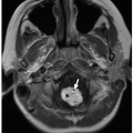
Radiographs and CT may show focal osteopenia, cortical irregularity, periosteal reaction, and contiguous soft tissue swelling. Findings are apparent on radiographs only after 7 to 10 days. Cortical irregularity and fluid adjacent to the bone and within the joint can be seen with US. Associated soft tissue changes on US include increased subcutaneous echogenicity, loss of normal soft tissue architecture, and reticular anechoic subcutaneous edema.54,55 US can be used to identify and guide drainage of superficial abscesses, which appear as hypoechoic or anechoic collections with posterior acoustic enhancement and peripheral hyperemia. Deep infection is better evaluated with CT or MRI. Although CT is superior at demonstrating osseous erosion, MRI best depicts the early changes of osteomyelitis. Abnormal marrow edema is evidenced by high T2-weighted and low T1-weighted signal intensity. Intravenous contrast may clarify regions of abscess formation (Fig. 12.15). Bone scintigraphy is sensitive for early detection but lacks anatomic detail.


TABLE 12.5 Differential Diagnosis of Aggressive-Appearing Pediatric Chest Wall Lesions


Underlying Etiologies


Disorders


Infection


Bacterial and fungal osteomyelitis


Benign soft tissue neoplasm


Plexiform neurofibroma


Benign osseous neoplasm


Osteoblastoma, fibrous dysplasia, and Langerhans cell histiocytosis


Malignant osseous neoplasm


Ewing sarcoma/primitive neuroectodermal tumor, osteosarcoma, and plasmacytoma


Metastasis


Neuroblastoma, rhabdomyosarcoma, leukemia, and lymphoma


Trauma


Subacute healing pathologic fracture in a benign osseous lesion


Vascular malformation


Kaposiform hemangioendothelioma


FIGURE 12.15 Staphylococcus aureus abscess and osteomyelitis in a 10-year-old boy with leukemia and disseminated infection. Axial T2-weighted MR image (top left), T1-weighted postcontrast fat-saturated MR image (bottom left), diffusion-weighted MR image (top right), and fused PET-MR image (bottom right) show a focal collection (arrows) with high T2 signal, peripheral enhancement, restricted diffusion, and hypermetabolic activity at the left anterior sixth costochondral junction with abnormal signal in the adjacent rib and soft tissues. A smaller collection is present on the right. Abnormal marrow signal in the spine was related to leukemic involvement.

Treatment consists of intravenous antibiotics and drainage of focal fluid collections.


Tuberculosis

Chest wall infection is a relatively rare manifestation of childhood tuberculosis. However, it should be considered in children with undiagnosed chest wall lesions, especially in endemic areas. The potential underlying mechanisms of chest wall tuberculous infection include direct extension from underlying pulmonary or pleural disease, direct extension from lymphadenitis of the chest wall, direct skin inoculation, and hematogenous dissemination. Tuberculous infection is usually more insidious than pyogenic bacterial infection. The presence of soft tissue calcification, abscess, osteolysis, and sequestra is suggestive56,57 (Fig. 12.16).


Actinomycosis

Actinomycosis results in a chronic granulomatous infection in immunocompromised pediatric patients. The thorax is affected in ˜15% of cases.58 Chest wall infection usually extends from pulmonary disease. Culture is negative in more than 50%; biopsy is frequently necessary for diagnosis.59


FIGURE 12.16 Pulmonary tuberculosis with sternal osteomyelitis and abscess in a 15-year-old boy. Axial enhanced lung window CT image (top) shows nodular and tree-in-bud pattern opacities with areas of cavitation. Axial enhanced soft tissue window CT image (middle) shows partially calcified subcarinal lymph nodes (arrow) and sternal destruction with surrounding soft tissue thickening and fluid collections. Sagittal bone window CT image (bottom) more clearly demonstrates the sternal osteolysis with adjacent sclerosis and cortical thickening.


FIGURE 12.17 Empyema necessitates in a 1-year-old girl with a history of gastropleural fistula and empyema. Coronal enhanced soft tissue window CT image shows a left pleural collection with surrounding pleural thickening and enhancement. The collection extends laterally through the eighth intercostal space into the subcutaneous tissues of the left lateral chest wall. There is significant atelectasis of the left lung. Pleural fluid culture showed multimicrobial infection.


Empyema Necessitates

Rarely, pleural empyema may directly extend into the soft tissues of the chest wall, usually anteriorly, resulting in empyema necessitates (Fig. 12.17). This is most commonly due to Mycobacterium tuberculosis and Actinomyces species.60


Neoplastic Disorders


Benign Neoplasms


Soft Tissue Tumors


Lipoma

Lipomas are proliferations of mature adipose tissue most commonly found in the upper back, neck, extremities, and abdomen.61 Other than causing a focal mass, they are usually asymptomatic. Grossly, they are soft, yellow, lobulated, and variably encapsulated. Microscopically, they consist of mature adipose tissue and variably prominent fibrous septa (Fig. 12.18). Genetically, a large proportion are characterized by dysregulation of the HMGA2 gene on the long arm of chromosome 12.

Radiographs may reveal a fat density mass without osseous erosion (Table 12.4). US shows an echogenic avascular mass with circumscribed or imperceptible borders.55 Homogenous fat attenuation (-100 Hounsfield units) is characteristic on CT (Fig. 12.19). MRI signal follows that of subcutaneous fat; fat-saturation technique can be applied for confirmation. A thin, fibrous capsule may surround subcutaneous and intermuscular lipomas but is usually absent in intramuscular lipomas, which tend to be infiltrative.62 Thin septa may be visible. Minimal or no enhancement is present. Fat necrosis can cause heterogeneity, in which case differentiation from liposarcoma cannot be definitively made, and biopsy is necessary.63


FIGURE 12.18 Lipoma in a 13-year-old girl. This 4 cm lesion resected from the chest subcutis consists grossly of pale yellow lobulated adipose tissue (left). Microscopically, the adipose tissue is traversed by thin septa containing mature fibroblasts (right, hematoxylin and eosin, original magnification, 100×).

Symptomatic lesions can be excised. Newer surgical techniques such as axillary approach subcutaneous endoscopic excision minimize scarring.64


Lipoblastoma/Lipoblastomatosis

In contrast to lipomas, lipoblastomas contain variable numbers of immature fat cells (lipoblasts), often with a myxoid extracellular matrix and highly vascular septa. The myxoid change can impart a shiny gelatinous quality to the gross specimen. However, variability in the maturity of lipoblasts can make lipoblastomas difficult to distinguish from lipoma, both grossly and microscopically (Fig. 12.20). The term “lipoblastomatosis” may be used for a lipoblastoma that exhibits diffuse involvement. Chromosomal rearrangements involving the PLAG1 gene are characteristic.65 Lipoblastomas present in infants and young children as painless masses in the extremities or trunk.62 They may recur after excision.


FIGURE 12.19 Lipoma in a 4-year-old boy who presented with right sided chest discomfort. Coronal enhanced CT image shows a well-defined, homogeneous fat attenuation mass (arrow) along the right lateral chest that extending into the fourth and fifth intercostal space.

Imaging features reflect the amount of fat within the lesion. Radiographs may demonstrate a fat and soft tissue
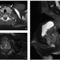
density mass. CT and MRI appearance is heterogeneous with fatty and nonfatty elements and occasionally a combination of nonenhancing cystic areas and enhancing soft tissue66 (Fig. 12.21).


FIGURE 12.20 Lipoblastoma in a 5-year-old boy. There was longstanding diffuse involvement (lipoblastomatosis) of the chest wall, axilla, and brachial plexus. Here, the features are of a “mature” lipoblastoma with adipose tissue, prominent fibrous septa, and only rare lipoblasts (hematoxylin and eosin, original magnification, 200x).

Definitive diagnosis of lipoblastoma requires biopsy and subsequent pathologic evaluation.


Desmoid Fibromatosis

Fibromatosis refers to a proliferation of fibroblasts or myofibroblasts, which tends to invade surrounding tissue and recur after incomplete excision. Desmoid fibromatosis (desmoid tumor) is the most common type of fibromatosis and presents as a painless mass in infants or young children, more commonly in males. It occurs in the deep or superficial soft tissue, most commonly in the trunk or extremities. Thoracic desmoids are usually isolated entities, whereas intra-abdominal desmoids are more frequently associated with Gardner syndrome. Young age and large tumor size are associated with increased risk of recurrence.67,68 Grossly, desmoid fibromatosis is firm, white gray and fairly well circumscribed, but not encapsulated. Microscopically, it shows plump active-appearing fibroblasts in fascicles or sheets68 (Fig. 12.22).


FIGURE 12.21 Lipoblastoma in a 12-year-old girl who presented with posterior chest wall deformity. Axial enhanced CT image (top), T1-weighted MR image (middle), and T1-weighted fat-saturated MR image (bottom) demonstrate a predominantly fatty lesion insinuating throughout paraspinal musculature, axilla, and intercostal regions bilaterally.

A nonspecific soft tissue mass is seen on radiography, often with erosion and deformity of adjacent bone (Fig. 12.23A). The attenuation on contrast-enhanced CT is usually greater than that of skeletal muscle. MRI usually shows homogeneous intermediate or low T1 and variable T2 signal intensity. Collagen-rich regions appear as curvilinear areas of low signal intensity on T2-weighted MR images (Fig. 12.23B). Enhancement after contrast is moderate to marked.69 Both an infiltrative and well-circumscribed pattern is seen in the pediatric population.70

Management is complex. Surgical resection has been a traditional approach. Adjuvant systemic chemotherapy and radiation may be used for rapidly growing symptomatic lesions. Due to treatment-associated morbidity, watchful waiting may be appropriate initial management. Recent study shows event-free survival does not significantly differ in children undergoing a period of observation compared to patients receiving surgical resection or systemic therapy.71


Myofibroma/Myofibromatosis

Myofibroma is a proliferation of fibroblasts and myofibroblasts that usually presents before 2 years of age with firm fleshy nodules. Though rare, it is the most common fibrous tumor of infancy. Three forms are described: solitary myofibroma, multicentric myofibroma in soft tissues, and multicentric myofibroma with visceral involvement. Solitary and multicentric myofibromas (myofibromatosis) typically occur in the head and neck, followed by the trunk and extremities. Lesions may increase in size and number or regress and even resolve. Nonvisceral lesions can be found in skin, subcutaneous tissue, muscle, or bone and have an excellent prognosis; however, reported mortality is up to 70% with visceral
involvement, which is most common in the lungs, gastrointestinal tract, and heart.68,72,73 Grossly, myofibroma/myofibromatosis is a firm white mass and variably well circumscribed. Microscopically, whorled bundles of fibroblastic cells are seen. A characteristic feature is the involvement of vessel walls (Fig. 12.24).

Only gold members can continue reading. Log In or Register to continue

Stay updated, free articles. Join our Telegram channel

Oct 13, 2018 | Posted by in PEDIATRIC IMAGING | Comments Off on Chest Wall

Full access? Get Clinical Tree

Get Clinical Tree app for offline access