Imaging: The plain x-ray is usually diagnostic. Typically, OS starts intramedullary but breaches the cortex and expands in the soft tissues. It is usually a combination of radiolucency and radiodensity, sometimes it is entirely eburneous with edges always faded. The pure osteolytic form is typical of the telangiectatic variety. The tumor soft tissue extension shows irregular, cloud-like radiodensities and/or stripes of density perpendicular to the cortex (sunray image). Occasionally, it is purely radiolucent, and it can be appreciated only by CT and MRI. At the periphery of the area where the tumor breaches the cortex, a triangular buttress of immature bone (Codman’s triangle) is seen. This is due to reactive bone acutely produced by the periosteum. Isotope bone scan is intensely hot even beyond the radiographic limits of the tumor. Rarely, it may reveal skips or distant bone mets. CT demonstrates intraosseous and extraosseous extension of the tumor and intratumoral radiodensities. With MRI, most OS exhibit the usual pattern of low T1 and high T2 signal. Sclerosing OS, however, have a low intensity on both T1 and T2 images. MRI is the best way to determine medullary tumor borders, epiphyseal invasion, and skip mets. Contrasted CT and MRI show the relationship with vessels, but in some cases, an angiogram may be more reliable. Patient workup always includes CT of the lungs where mets may appear as radiodense round nodules.
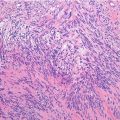
Histopathology: OS is fleshy soft in cellular areas with little matrix, firm and rubbery in fibroblastic areas with collagen production, gritty to stone-hard in osteogenic areas, and cartilaginous to myxoid in chondroblastic areas. Hemorrhage, necrosis, and cystic alterations are common. The most relevant diagnostic feature is constant permeation of marrow spaces, trapping host trabecular bone along its margins, predominantly. Cortex is also permeated and usually breached. An endosteal and periosteal production of reactive bone is associated. OS may invade the joint capsule and ligaments. Rarely, OS plugs are found in the adjacent veins. Skip mets, usually in the same but also in the adjacent cross-joint bone, may be detected in a small percentage of cases. Microscopically OS has a wide range of histological presentations but the characterizing feature is represented by high-grade sarcomatous cells producing osteoid and woven bone. The less osteogenic areas of the tumor (usually the periphery) are highly cellular and show more clear-cut features of high-grade malignancy as compared to more osteoid or bone-rich central areas of the tumor. Cells are large, with striking pleomorphism, hyperchromia, prominent nucleoli, and frequent atypical mitoses, although some 10 % of cases may show little anaplasia and lead to confusion with benign entities such as osteoblastoma, chondroblastoma, and giant cell tumors. Tumor osseous matrix varies from slender lacelike seams of osteoid to islands or dense sheets of woven bone. No regular trabeculae rimmed by osteoblasts are produced by OS cells. Where OS is intensely sclerotic, cells are scarce and small, with no mitoses; tissue is scarcely vascular and may be necrotic. In these areas, features of malignancy may be absent and diagnosis of OS is suggested by the permeative pattern of the tumor. Occasionally, OS is extensively chondroblastic (as a high-grade chondrosarcoma) or fibroblastic (similar to a fibrosarcoma). The osteoid-osseous production, which identifies the OS, may be found only in the microscopic study of the entire specimen. Reactive giant cells are seen, particularly in areas of hemorrhage. Particularly at the periphery of the tumor, reactive osteogenesis associates and should not be confused with tumorous osteogenesis. Histochemical stains show a high content of alkaline phosphatase in OS cells.
Related posts:


Stay updated, free articles. Join our Telegram channel

Full access? Get Clinical Tree


