Fig. 8.1
This is the chest x-ray of an occult pneumothorax. This image belongs to the same patient in Video 8.9
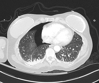
Fig. 8.2
This CT scan shows the pneumothorax not to be as insignificant as previously believed to be on the x-ray. The image belongs to the same patient in Fig. 8.1 and Video 8.9
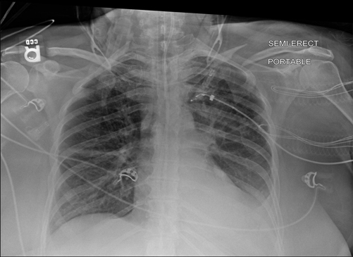
Fig. 8.3
This is a chest x-ray showing pleural effusion. It belongs to the same patient on Video 8.10. The ultrasound video shows how a large pleural effusion can look moderate or small on portable x-ray
Summary
Ultrasound has many applications, especially when evaluating a patient whose clinical status is deteriorating. Expertise is required to trust the examination in these late stages of resuscitation. Therefore, previous training and experience in performing this test in healthier individuals will come in handy.
Appendix
Video 8.1 A flat IVC. In the setting of hypotension this image is diagnostic of hypovolemia.
Video 8.2 A hyperdynamic heart. In the setting of hypotension, a hyperdynamic heart should be considered a sign of hypovolemia.
Video 8.3 Positive EFAST in Morrison’s pouch. On a hypotensive trauma patient, this fluid is blood until proven otherwise.
Video 8.4 Positive EFAST for intrathoracic fluid. On a hypotensive trauma patient, this fluid is blood until proven otherwise.
Video 8.5 Poor contractility.
Video 8.6 Right-sided cardiac dysfunction.
Video 8.7 Cardiac tamponade. Notice the complete compression of the right-sided cardiac structures.
Video 8.8 This video shows a pericardial effusion and a pleural effusion. In this case it was an injury to the right ventricle decompressing into the thoracic cavity.
Video 8.9 Pneumothorax. This video shows the lung point sign demarcating the penumothorax.
Video 8.10 Pleural effusion.
References
1.
2.
3.
4.
5.
Abbasi S, Farsi D, Hafezimoghadam P, Fathi M, Zare MA. Accuracy of emergency physician-performed ultrasound in detecting traumatic pneumothorax after a 2-h training course. Eur J Emerg Med. 2012.
6.
7.
8.
9.
10.
11.

