, Valery Kornienko2 and Igor Pronin2
(1)
N.N. Blockhin Russian Cancer Research Center, Moscow, Russia
(2)
N.N. Burdenko National Scientific and Practical Center for Neurosurgery, Moscow, Russia
Primary lymphomas (PCNSL). Until recently, primary lymphoma of the central nervous system has been quite a rare disease and accounted for about 1% of all brain tumors. Among other diseases, in which the risk of primary CNS lymphoma increases, are systemic collagenoses (systemic lupus erythematosus, rheumatoid arthritis, etc.) and chronic viral infections, in particular, Epstein-Barr virus infection. By their histological structure, PCNSLs are virtually always represented by a non-Hodgkin’s type and are mostly B-cell lymphomas (Bergmann and Edel 1991). Primary T-cell CNS lymphomas are also described but are rare. By their location, 90% of all lymphomas are supratentorial. Majority of all cases (73%) are presented with a solitary lesion; in other cases multiple brain lesions are determined. Leptomeningeal or dural distribution typically occurs in systemic lymphoma.
In order to verify the histological diagnosis and for subsequent chemotherapy and radiation therapy for suspected brain lymphoma, a stereotactic biopsy (STB) is advisable. Therefore, establishing the diagnosis of primary lymphoma at the preoperative stage is an extremely important task, which would significantly reduce the percentage of unjustified surgical interventions.
Tumor lesions look like quite clearly demarcated, space-occupying lesions with various sizes, often rounded, with clear contours, so they may mimic metastases or glioblastoma. At the same time, a solid, compact tumor structure is observed in some patients and the presence in the stroma of necrotic areas with different sizes in others (a smaller proportion). The latter is considered a typical manifestation of lymphomas in HIV-infected patients.
According to CT, the tumor in most cases has an increased density. An isodense tumor is diagnosed in only 20% of cases. A necrotic area in the center of the tumor has lower density relative to the stroma. Intense accumulation of the contrast medium in the stroma of lymphoma after intravenous injection is the characteristic CT manifestation of the tumor and occurs almost always. Edema of the surrounding brain substance, with varying severity, is identified as an area with decreased density around a hyperdense tumor lesion (Fig. 30.1).
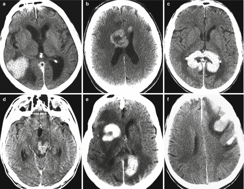
Fig. 30.1
Lymphoma. CT variants of lymphomas with different structure and location: (a) solid tumor in the right temporoparietal convexital area; (b) mixed lymphoma with central necrosis of the genus of corpus callosum; (c) solid tumor of the splenium of the corpus callosum; (d) nodular solid tumor of the midbrain; (e) mixed, multiple lymphoma; (f) multiple, convexital tumor with a hemorrhage
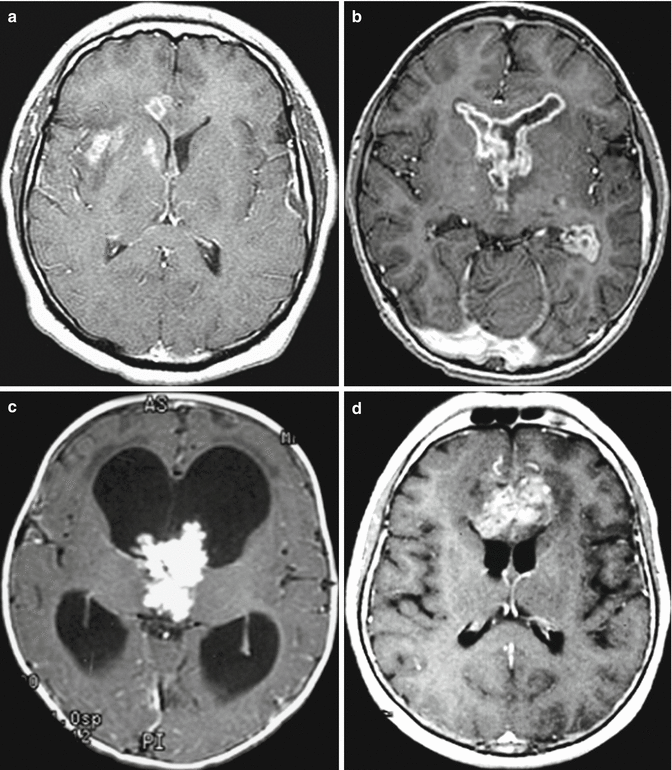
The lymphoma stroma on standard MRI sequences has an isointense or slightly hypointense MR signal relative to the unaffected gray matter. In the presence of a necrotic decay cavity in the tumor (usually in the central sections of the tumor), this area is characterized by a high and low signal in T2-weighted and T1-weighted images, respectively. Perifocal edema with a high signal on T2-weighted MRI and a low signal on T1-weighted MRI was noted in 30–40% of cases. With intravenous administration of the contrast medium, intense homogeneous opacification of the tumor stroma is observed, in the absence of accumulation in the necrotic area (if any)—ring-shaped contrast enhancement (Figs. 30.2, 30.3, and 30.4).
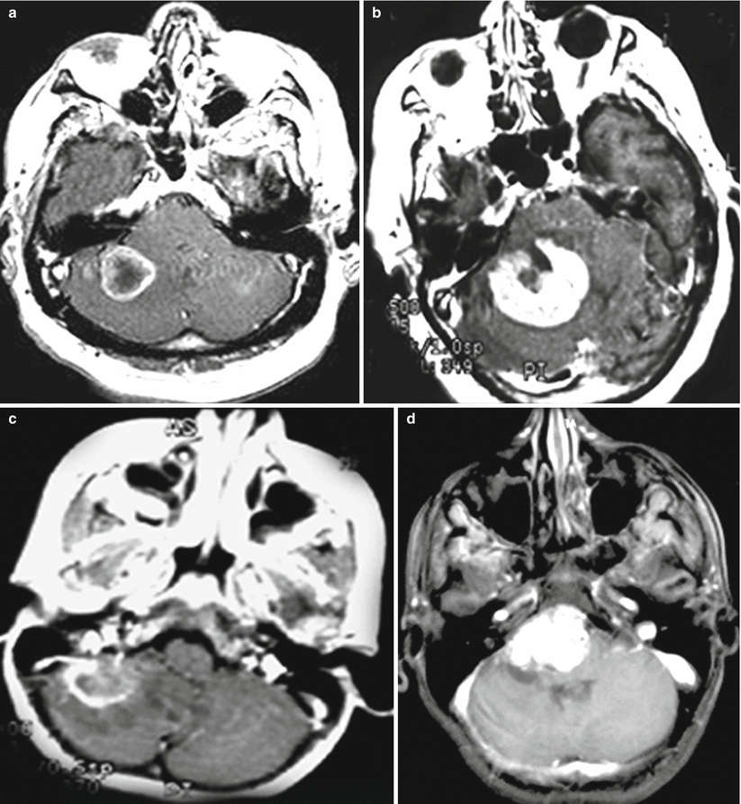
Fig. 30.2
Space-occupying lesions of the cerebellopontine angle. T1-weighted MRI with contrast enhancement. The differential diagnosis of a variety of lesions characterized by intense accumulation of the contrast agent and dislocation of the brainstem structures: lung cancer metastasis (a), lymphoma (b), ependymoma (c), hemangioblastoma (d)