Collagen-vascular Diseases


Collagen-vascular Diseases


W. Richard Webb


Collagen-vascular diseases (CVD) may be associated with focal or diffuse pulmonary abnormalities. The type and frequency of lung abnormalities vary with the specific disease. The two most common conditions associated with lung disease are rheumatoid arthritis (RA) and scleroderma (progressive systemic sclerosis [PSS]).

Most CVD can cause chronic interstitial pneumonia with clinical, radiographic, high-resolution CT (HRCT), and pathologic features indistinguishable from those of the idiopathic interstitial pneumonias (see Chapter 13). CVD may result in usual interstitial pneumonia (UIP), nonspecific interstitial pneumonia (NSIP), lymphoid interstitial pneumonia (LIP), and organizing pneumonia (OP), also known as bronchiolitis obliterans OP (BOOP). Individual CVD may also be associated with specific types of lung abnormality.


RHEUMATOID ARTHRITIS

Rheumatoid arthritis (RA) is commonly associated with thoracic abnormalities, including interstitial pneumonia and fibrosis (UIP and NSIP are common), pleural effusion or pleural thickening, necrobiotic nodules, OP, bronchiectasis, and bronchiolitis obliterans (Table 14-1).

Clinical evidence of arthritis precedes the development of pulmonary or pleural disease in about 90% of patients, and 90% have a positive serum rheumatoid factor. Although RA is twice as common in women, extraarticular manifestations of RA, including lung disease, are more common in men. Radiographic findings vary with the manifestation of RA present (see Table 14-1).


Pleural Disease

Pleural disease, either pleural effusion or pleural thickening, is common in patients with RA being seen in up to 40% of patients at autopsy. However, radiographic evidence of pleural thickening or pleural effusion is present in only 5% to 20% of patients, and it often is asymptomatic. Pleural effusions are usually small and unilateral, being more common on the right. Generally, effusion is not associated with radiographic evidence of lung disease. On HRCT, pleural effusion is visible more often than on radiographs and is seen in more than 40% of patients with findings of interstitial pneumonia.


Interstitial Pneumonia and Fibrosis

Interstitial pneumonia and fibrosis, the most common pulmonary manifestation of RA, is most common in middle-aged men. Dyspnea is common. Histologically, it may represent either NSIP or UIP.

Although pulmonary function abnormalities are present in up to 40% of patients with RA, only about 5% to 10% of patients with RA have radiologically detectable interstitial disease.

On HRCT, interstitial pneumonia may be associated with a spectrum of abnormalities ranging from ground-glass
opacity to fine reticulation or coarse reticulation with honeycombing. Findings of pulmonary fibrosis (irregular reticulation, traction bronchiectasis, honeycombing) are seen on HRCT in about 10% of patients; ground-glass opacity is seen in 15%.


TABLE 14.1 Rheumatoid Arthritis


Arthritis precedes lung disease in 90%


Lung disease more common in men


Pleural effusion or pleural thickening in 5%-20%


Interstitial pneumonia 10%-25%


Histologic pattern may be UIP, NSIP OP/BOOP, or LIP


Ground-glass, reticulation, honeycombing


Reticulation usually finer than IPF


Basal and peripheral predominance


Immediate subpleural sparing may be present (atypical with IPF)


Anterior upper lobe honeycombing (atypical with IPF)


Consolidation with OP/BOOP


Rheumatoid nodules


Peripheral, well-defined


Caplan’s syndrome


Bronchiectasis in 20%


Bronchiolitis obliterans rare


Lymphoid interstitial pneumonia or follicular bronchiolitis


Cystic appearance


Centrilobular nodules


Pulmonary hypertension


IPF, idiopathic pulmonary fibrosis; OP/BOOP, organizing pneumonia/bronchiolitis obliterans organizing pneumonia.


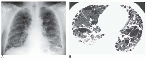
FIG. 14.1. Pulmonary fibrosis with honeycombing in a man with rheumatoid arthritis. A: Chest radiograph shows a coarse reticular pattern typical of honeycombing. Lung volume is reduced. B: Extensive honeycombing is visible on HRCT. This is a usual interstitial pneumonia pattern of lung disease. A right pleural effusion is also present.

The appearance of RA with interstitial fibrosis may be indistinguishable from that of idiopathic pulmonary fibrosis (IPF) (Figs. 14-1 and 14-2). However, because some cases are associated with the histologic pattern of NSIP (see Chapter 13), reticular opacities visible in RA tend to be finer than those seen with IPF (Figs. 14-3 and 14-4), and honeycombing is less common and less severe. Also, anterior upper lobe honeycombing appears to be more common in the CVD than in IPF (see Figs. 14-2B and 14-4B). In some patients, this may be a manifestation of LIP rather than UIP or NSIP.


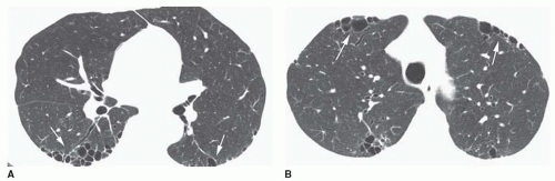
FIG. 14.2. Pulmonary fibrosis with honeycombing in a woman with rheumatoid arthritis. A: Patchy subpleural honeycombing (arrows) is visible in the posterior lung. B: Patchy honeycombing is also present in the upper lobes. Anterior upper lobe honeycombing (arrows) is more common in collagen-vascular diseases than in idiopathic pulmonary fibrosis. This may represent usual interstitial pneumonia or lymphoid interstitial pneumonia.

Predominance in the posterior and subpleural lung and at the lung bases is typical of reticular opacities and ground-glass opacity (Fig. 14-5). Relative sparing of the immediate subpleural lung may be seen, a finding associated with the histologic pattern of NSIP (see Fig. 14-4).

OP/BOOP may also occur in patients with RA, having typical radiographic and CT features of patchy consolidation or nodules (see Chapter 13).


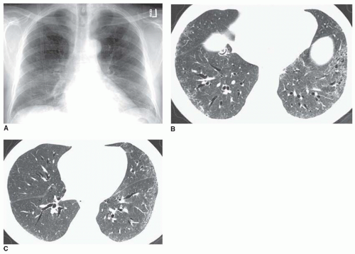
FIG. 14.3. Pulmonary fibrosis in rheumatoid arthritis with reticular opacities. A: Chest radiograph shows some reduction in lung volume and a mild nonspecific reticular abnormality at the lung bases. B: Fine reticular opacities are visible at the lung bases with evidence of traction bronchiectasis. There is no evidence of honeycombing. C: At a higher level, reticular opacities are less severe and predominate on the left. A distinct subpleural and lower lobe predominance is visible. This may be termed a nonspecific interstitial pneumonia or usual interstitial pneumonia pattern.


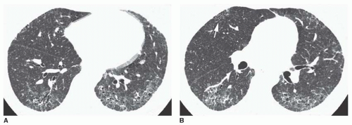
FIG. 14.4. Rheumatoid arthritis with reticular opacities and subpleural sparing. A: Fine reticular opacities are visible at the lung bases with evidence of traction bronchiectasis. Although there is a predominance in the subpleural lung, the immediate subpleural lung is less severely involved. This appearance tends to be associated with the histologic pattern of nonspecific interstitial pneumonia. B: At a higher level, subpleural sparing is also visible. Despite the absence of lower lobe honeycombing, patchy honeycombing is visible in the anterior upper lobe (arrow). This finding suggests a collagen-vascular disease.


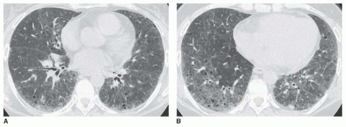
FIG. 14.5. Ground-glass opacity in a woman with rheumatoid arthritis. A: HRCT shows patchy ground-glass opacity with a subpleural predominance. B: The abnormality is more extensive at the lung bases. This appearance may reflect nonspecific interstitial pneumonia or lymphoid interstitial pneumonia (LIP). Small cystic lucencies are visible within the abnormal lung. These may represent traction bronchiectasis associated with lung fibrosis or cysts associated with LIP.


Rheumatoid Nodules

Rheumatoid (necrobiotic) nodules are an uncommon manifestation of RA. Their histology is identical to that of subcutaneous nodules. They are often asymptomatic but tend to appear and disappear in conjunction with subcutaneous nodules. They range in size from a few millimeters to 5 cm or more and may be solitary or multiple and numerous. Rheumatoid nodules predominate in the lung periphery and are typically well-defined (Fig. 14-6; see also Fig. 9-49 in Chapter 9). They may cavitate, having thick walls that become thin with healing. Pleural effusion may be associated, and cavitary nodules in the periphery may lead to pneumothorax.


Caplan’s Syndrome

Caplan’s syndrome is a rare manifestation of RA occurring in coal miners or patients with occupational exposures to silicates. Nodules are present that resemble necrobiotic nodules histologically but are characterized by concentric layers of light collagen and black dust-laden cells. Caplan’s syndrome is characterized by single or multiple lung nodules ranging from a few millimeters to 5 cm in diameter, similar to rheumatoid nodules. The nodules may cavitate. An upper lobe predominance may be seen, resembling silicosis or coal-worker’s pneumoconiosis, but nodules in Caplan’s syndrome appear rapidly in “crops” in contrary to the slow progression of silicosis.


Bronchiectasis

Bronchiectasis is associated with RA, particularly in smokers. It may reflect chronic infection, which has an increased incidence in rheumatoid patients or, associated with i nfection, it may be a factor in the development of RA. Also, bronchiectasis is commonly present in patients with bronchiolitis obliterans (Fig. 14-7). On CT, it is present in 20% of patients.


Bronchiolitis Obliterans

Bronchiolitis obliterans (see Chapter 23) is a rare manifestation of RA. Although it is often associated with penicillamine treatment, this is not always the case. Radiographs
are normal or show increased lung volumes. HRCT may show bronchiectasis, mosaic perfusion, and air trapping (see Fig. 14-7).


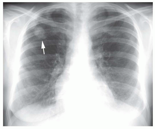
FIG. 14.6. Rheumatoid nodule. A young man with rheumatoid arthritis shows a well-defined nodule in the peripheral right upper lobe. Small pleural effusions are also present (arrow).


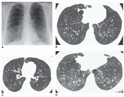
FIG. 14.7. Bronchiectasis and bronchiolitis obliterans in a young woman with rheumatoid arthritis. A: Chest radiograph shows large lung volume. B and C: HRCT scans at two levels show extensive bronchiectasis and regional differences in lung attenuation due to mosaic perfusion. D: Postexpiratory HRCT at the same level as (B) shows patchy air trapping typical of bronchiolitis obliterans.


Follicular Bronchiolitis and Lymphoid Interstitial Pneumonia

Follicular bronchiolitis is also a rare airway manifestation of RA; it is characterized by dyspnea. A lymphocytic infiltrate in the walls of small airways is present histologically. Radiographs may show a reticular or reticulonodular pattern. HRCT shows small centrilobular nodules or tree-in-bud. More extensive lymphocytic infiltrations may be associated with findings of LIP such as ground-glass opacity or lung cysts (see Fig. 14-5; see also Figs. 13-32 and 13-35 in Chapter 13).


Pulmonary Hypertension

Pulmonary hypertension usually occurs secondary to pulmonary fibrosis. Rarely it develops in the absence of lung disease, perhaps related to vasculitis.


PROGRESSIVE SYSTEMIC SCLEROSIS (SCLERODERMA)

PSS, also known as scleroderma, is a generalized disease of connective tissue often associated with vasculitis. Women outnumber men by three to one. Only 1% present with pulmonary symptoms, but up to 75% have evidence of pulmonary disease at some point in their disease course.

PSS leads to some degree of interstitial fibrosis in nearly all patients. PSS is commonly associated with NSIP, UIP, pulmonary vasculitis, and pulmonary hypertension (Table 14-2).


Interstitial Pneumonia and Fibrosis

As with RA, chest radiographs may appear normal despite abnormal pulmonary function tests. The incidence of radiographically recognizable interstitial disease is about 25%. Chest radiographs may show a reticular abnormality with lower lobe predominance indistinguishable from that of IPF.
In early cases, ill-defined ground-glass opacity may be seen in the bases (Fig. 14-8A). With progression, reticulation may progress from fine to coarse, associated with progressive loss of lung volume.


TABLE 14.2 Scleroderma


Lung disease in 75%


More common in women


Interstitial pneumonia


Histologic pattern may be NSIP, UIP, OP (BOOP)


Ground-glass, reticulation, honeycombing


HRCT appearances similar to rheumatoid arthritis


Pleural effusion or thickening in 35%


Esophageal dilatation in 40%-80%


Mediastinal lymph node enlargement in 60%

Only gold members can continue reading. Log In or Register to continue

Stay updated, free articles. Join our Telegram channel

Oct 10, 2016 | Posted by in CARDIOVASCULAR IMAGING | Comments Off on Collagen-vascular Diseases

Full access? Get Clinical Tree

Get Clinical Tree app for offline access