Congenital Brain Malformations


Congenital Brain Malformations


Teresa Chapman, MD


INTRODUCTION

Development of the brain is, needless to say, an extraordinarily complex process. Organization of the primitive brain begins within the first 20 days of embryonic development and continues postnatally. The neural plate is a longitudinal collection of precursor cells for the brain and spine. By 3 weeks of development, three distinct broadenings reflecting the primary brain vesicles are already apparent: the prosencephalon (forebrain precursor), the mesencephalon (midbrain), and the rhombencephalon (hindbrain). During the 4th week, a process called neurulation converts the neural plate to a hollow neural tube. The neural tube undergoes rapid elongation, closure, and cephalic folding. The brainstem anatomy and closure of the fourth ventricle is established early, whereas development of the cerebellum and cerebral hemispheres continues through gestation and into postnatal life. Most brain cells arise from neuroepithelium lining the neural canal. The elegant layering of nerve cells in the cerebral cortex is formed by a stepwise proliferation of progenitor cells within the germinal matrix along the margins of the lateral ventricles, followed by outward migration along radially projecting glial fibers. Similarly, the cerebellar cortices develop by outward migration of neuroblast precursor cells generated by an internal germinal layer adjacent to the ventricular zone.1 Failure of any of the innumerable steps required for these processes—neural tube closure, folding, induction of neuroepithelial migration, and differentiation— can result in a congenital brain malformation. In this chapter, the most commonly encountered congenital brain malformations will be described.


SUPRATENTORIAL MALFORMATIONS


Disorders of the corpus callosum

The corpus callosum is a large midline bundle of crossing white matter fibers connecting the cerebral hemispheres. It develops from approximately 8 weeks through 18 weeks of fetal gestation. The corpus callosum grows primarily from anterior to posterior, with the genu forming first, followed by the anterior body, posterior body, and splenium. The rostrum, however, forms after the splenium, between 18 and 20 weeks of gestational age (Fig. 30.1).2 Partial or complete malformation (agenesis) of the corpus callosum has an incidence of 1 in 4,000 live births.3 Although agenesis of the corpus callosum can be an isolated finding, callosal malformations are more commonly seen in conjunction with other brain anomalies, including Chiari malformation, encephalocele, migrational anomalies, holoprosencephaly (HPE), and malformations of the brainstem or cerebellum.3,4 The prognosis depends on all associated abnormalities and underlying genetic influences. Although isolated callosal malformations may be asymptomatic, seizures, microcephaly, and developmental delays are frequently observed.5

Partial dysgenesis most commonly manifests with absence of the splenium and a portion of the mid- to posterior body (Fig. 30.2A, B). Diffuse or segmental callosal thinning may result secondary to a vascular or inflammatory insult that destroys white matter volume (Fig. 30.2C, D). A rare association with callosal agenesis or dysgenesis is a midline lipoma, which is presumed to arise from abnormal differentiation of the meningeal precursor cells.6 Of note, midline lipomas can be present even in

the setting of a normally formed corpus callosum, and they have no clinical implication (Fig. 30.3).


FIG. 30.1 • Corpus callosum anatomy on brain MRI of normal 5-year-old child. A: Sagittal T1-weighted MR image shows large hyperintense bundle of midline crossing myelinated fibers. Components of normal corpus callosum are annotated here. B: Axial T2-weighted MR image shows dark signal of myelinated fibers anteriorly and posteriorly (arrows).


FIG. 30.2 • Corpus callosum partial dysgenesis and thinning. Sagittal T1-weighted (A) and axial T2-weighted (B) MR images from a 4-year-old child with Chiari malformation and corpus callosal dysgenesis show partial absence of corpus callosum. Posterior body, splenium, and rostrum are absent (white arrows on A). Axial image shows absence of crossing fibers (black arrows in B) that should be readily apparent. Lateral ventricles assume a parallel orientation, typical of partial and complete corpus agenesis. Sagittal T1-weighted (C) and axial T2-weighted (D) MR images from 5-month-old infant with large left hemispheric porencephalic cyst (PC) show a fully formed but markedly thinned (white arrows) corpus callosum secondary to white matter volume loss. Note normal convergence of lateral ventricles toward one another on axial plane, in contrast to partial dysgenesis case.


FIG. 30.2 • (Continued).


Migrational abnormalities

Normal cerebral cortical development depends on neuronal cell proliferation, differentiation, migration, and organization.7, 8 and 9 Disruption of any of these processes may occur in the context of genetic anomalies, congenital infections such as cytomegalovirus or toxoplasmosis, ischemic injury, and toxin exposure or may occur sporadically. Cellular proliferation and migration may be either insufficient or exuberant, resulting in very different appearances of the brain.9 Table 30.1 summarizes the various malformations included within the category of migrational anomalies. Lissencephaly, or “smooth brain,” is a term that applies to a number
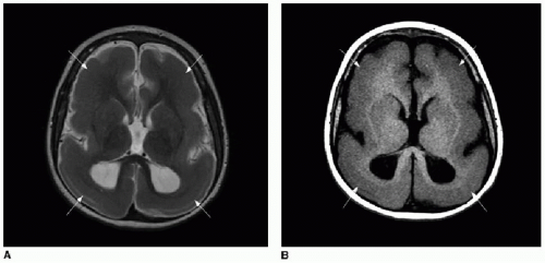
of diffuse proliferative or migrational disorders characterized by abnormal simplification of the cortical convolutions (Fig. 30.4).10,11 Agyria refers to absence of cortical gyri, whereas pachygyria refers to abnormally broad cerebral gyri; these terms also fall within the category of lissencephaly. A number of genetic defects causing lissencephaly have been identified.11 While diffuse migrational anomalies present with severe developmental delay, intractable seizures, and hypertonia, the focal malformations tend to present with medical refractory seizures, and we will focus on these entities here.


FIG. 30.3 • Interhemispheric/callosal lipoma. Sagittal T1-weighted MR image without (A) and with (B) fat saturation in a 10-year-old female shows fat signal in the midline, along the mid- and posterior corpus callosum (arrow), consistent with interhemispheric lipoma.


Table 30.1 CATEGORIZATION OF NEURONAL MIGRATION ANOMALIES


Pathogenesis


Malformation


Description


Disordered proliferation


Decreased


Microlissencephaly


Diffusely thin cerebral mantle, markedly abnormal sulcation


Increased


Hemimegalencephaly


Hamartomatous overgrowth of a cerebral hemisphere or lobe of hemisphere


Focally abnormal


Focal cortical dysplasia


Lack of normal cortical lamination, focal blurring of gray-white border


Disordered migration


Decreased


Classic lissencephaly


“Smooth brain”; marked thickening of cerebral cortex, absent formation of sylvian fissures; usually no other CNS malformation


Increased


Cobblestone lissencephaly


CNS manifestation of congenital muscular dystrophy; variable pachygyria and polymicrogyria, thickened cortex, focal interhemispheric fusion


Focally ectopic


Gray matter heterotopias


Nodular ectopic foci of disorganized epileptogenic neurons and glial cells


Disordered organization


Terminal


Polymicrogyria


Excessive cortical convolutions, most common near Sylvian fissures


Subependymal to terminal


Schizencephaly


Cerebral cleft from ventricle to pial surface; lined by malformed gray matter


From Rollins N. Congenital brain malformations. In: Coley BD. Caffey’s Pediatric Diagnostic Imaging. 12th ed. Philadelphia, PA: Elsevier Saunders: 2013;299-320.


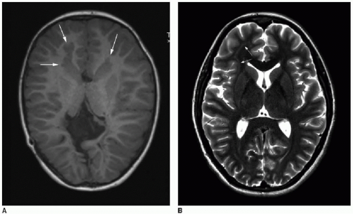
Gray matter heterotopias are nodules or masses of abnormal tissue that follow gray matter signal on MR sequences and are histologically characterized by a combination of normal neurons and glial cells.7,9 These may be found in the periventricular or subcortical white matter (Fig. 30.5). Periventricular heterotopias tend to manifest with focal seizures, whereas subcortical heterotopias more commonly are associated with partial complex and tonic-clonic seizures.7 The more extensive the heterotopia, the more likely the child will be affected by developmental delays in
addition to seizures.7 A diffuse form of gray matter heterotopia is called subcortical band heterotopia (SBH), wherein a thick band of tissue isointense to gray matter replaces a variable volume of white matter, and only a thin rim of white matter is evident between the heterotopic tissue and the cortex (Fig. 30.4). SBH falls into the category of lissencephaly, and its clinical severity depends on the extent of cortical abnormalities, band thickness, and ventricular enlargement.11


FIG. 30.4 • Lissencephaly. Axial (A) T2-weighted and T1-weighted (B) MR images of 7-year-old patient’s brain show markedly simplified cerebral sulcation pattern bilaterally. Widespread replacement of expected subcortical white matter signal is abnormally replaced by gray matter signal (arrows), consistent with diffuse subcortical band heterotopia.


FIG. 30.5 • Gray matter heterotopia. A: Periventricular gray matter heterotopia in bifrontal lobes (arrows) shown on axial T1-weighted MR image of a 5-year-old child with seizures. Note also complete agenesis of corpus callosum. B: Subcortical thin band of heterotopia (arrows) in right frontal white matter shown on axial T2-weighted MR image of 17-year-old patient with seizures. Signal of abnormal white matter nodules and band followed gray matter signal on all sequences (not shown here).

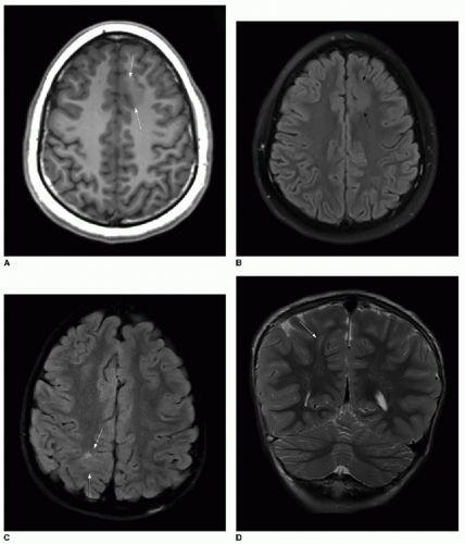
Focal cortical dysplasia (FCD) has recently been classified according to the histologic cortical laminar structure and architectural disruption, cell composition, and presence of associated destructive lesions.12 Types I and II are isolated lesions that are both characterized by abnormal cortical lamination. They are differentiated based on absence (type I) or presence (type II) of dysmorphic neurons. Type II dysplasias may also have eosinophilic balloon cells, which were first described in 1971 and may be grossly abnormal glial cells.13 Type III lesions are focal areas of cortical lamination abnormalities associated with a destructive lesion such as hippocampal sclerosis, glial tumor, vascular malformation, or a focal insult acquired in fetal or early life such as infection or infarction.12 Identification of FCD on brain MRI requires careful scrutiny of high-resolution images. The hallmarks of type I FCD include cortical thickening and loss of a distinct border between gray and white matter (Fig. 30.6A, B). Type II is more readily identified on MR. The transmantle sign describes a radially oriented linear or conical subcortical T2 hyperintensity, reflecting the radial extension of balloon cells and ectopic neurons from the cortex into the affected white matter (Fig. 30.6C, D). This sign can be useful in differentiating types I and II FCD.14 Surgical resection of FCD results in a seizure-free outcome in 33% to 75% of cases, depending on center-specific selection criteria for surgical eligibility.14,15

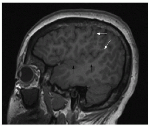
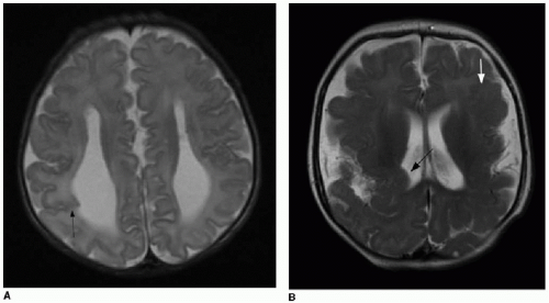
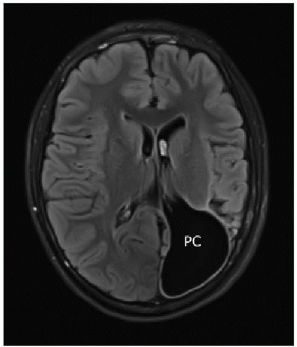
Polymicrogyria (PMG) accounts for approximately 20% of all cortical malformations observed. It is characterized by an excessive number of small cortical gyri, with distortion of the expected gyral and sulcal pattern.8,16 The extent of PMG varies greatly, as does the range of clinical manifestations. When PMG is bilateral, it is most often found along the Sylvian fissures (Fig. 30.7).17 Several chromosome abnormalities are associated with PMG; the most common are the 1p36.3 microdeletion and the 22q11.2 microdeletion, both of which are associated with unilateral or bilateral perisylvian PMG.17 Schizencephaly is a specific pattern of PMG defined as a full-thickness cleft in the brain.18,19 These clefts must be lined by PMG to be called schizencephaly (Fig. 30.8), as opposed to a porencephalic cyst (Figs. 30.2D and 30.9), which can be thought of as an intraparenchymal cerebrospinal fluid (CSF) space lined by white matter or gliosis, resulting presumably from a fetal insult causing focal brain destruction.17 Schizencephaly has been described as closed lip or open lip—if the cleft contains CSF density or signal, it is open lip, whereas closed-lip anomalies lack interposed CSF on imaging.18,19 However, differentiation of the two types is generally not clinically relevant. Neuronal migrational abnormalities can be associated with other brain malformations, so it is important to search carefully for other findings.


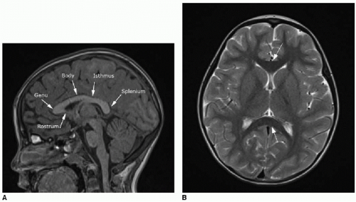
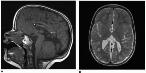
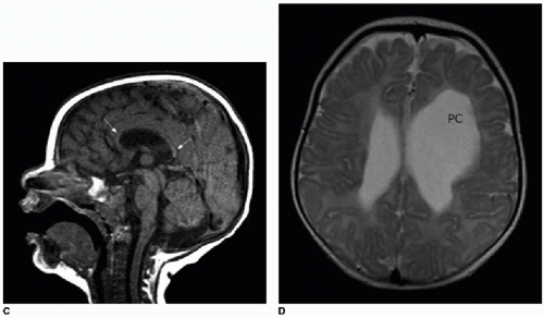
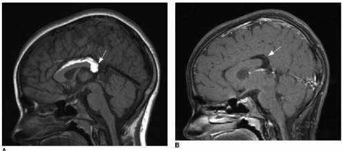
FIG. 30.6 • Focal cortical dysplasia. Type I FCD in 15-year-old patient with seizures is evident on T1-weighted (A) and T2-weighted (B) axial images as area with loss of gray-white matter differentiation and abnormal signal on both sequences (arrows). C: Type II FCD in 5-year-old patient manifests with hyperintense T2-weighted signal on fluid attenuation and inversion recovery (FLAIR) sequence in axial plane (arrows). D: Transmantle sign is apparent on coronal T2-weighted image, with high signal radiating from affected cortex toward the ventricular surface. Following surgical resection, histopathology confirmed presence of balloon cells.


FIG. 30.7 • Perisylvian polymicrogyria. Sagittal T1-weighted thinslice MR image of 16-year-old seizure patient’s brain shows excessive number of small cortical gyri at upper aspect of perisylvian region (white arrows), consistent with PMG. Contrast with normal gyral pattern along Sylvian fissure more inferiorly (black arrows).


FIG. 30.8 • Examples of schizencephaly. A: Axial T2-weighted brain MR image of male neonate with prenatal diagnosis of agenesis of corpus callosum and ventriculomegaly demonstrates abnormal gray matter-lined right parietal cleft coursing from cortical surface to ventricular surface (arrow). Note parallel configuration of lateral ventricles, typical of absent corpus callosum. B: Axial T2-weighted brain MR image of 5-month-old female infant at the level of lateral ventricles shows extensive malformation of cerebral hemispheres. Right parietal cleft anomaly creates indentation upon ependymal surface (black arrow), characteristic of schizencephaly. Dysplastic gray matter lines the cleft. Note also polymicrogyria in left frontal lobe (white arrow).


FIG. 30.9 • Porencephalic cyst. Axial T2-weighted image of the brain of 13-year-old shows large white matter-lined CSF space contiguous with left lateral ventricle. (PC, porencephalic cyst.)


Holoprosencephaly

HPE describes a spectrum of mild to severe brain malformations characterized by incomplete cleavage of the two cerebral hemispheres, occurring secondary to disruptions in the early induction of the forebrain (prosencephalon).1 Median facial anomalies, including hypotelorism, cyclopia (single eye), cebocephaly (single nostril), cleft palate, and single midline incisor, may be seen with the more severe forms of HPE, reflecting the closely related development of the face and the brain.1,20 HPE has been found to be associated with chromosome abnormalities, single gene disorders, and maternal factors including pre-existing diabetes, exposure to teratogenic agents such as alcohol, and lower educational level.21 The prevalence of HPE at birth is low, estimated to be 1.3 per 10,000 live births.22,

Only gold members can continue reading. Log In or Register to continue

Stay updated, free articles. Join our Telegram channel

May 24, 2016 | Posted by in PEDIATRIC IMAGING | Comments Off on Congenital Brain Malformations

Full access? Get Clinical Tree

Get Clinical Tree app for offline access