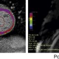
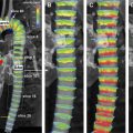
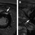
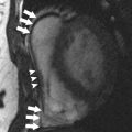
Magnetic resonance (MR) imaging of the coronary arteries has been challenging, owing to the small size of the vessels and the complex motion caused by cardiac contraction and respiration. Free-breathing, whole-heart coronary MR angiography has emerged as a method that can provide visualization of the entire coronary arterial tree within a single 3-dimensional acquisition. Although coronary MR angiography is noninvasive and without radiation exposure, acquisition of high-quality coronary images is operator dependent and is generally more difficult than computed tomographic angiography. This article explains how to optimize acquisition of coronary MR angiography for reliable assessment of coronary artery disease. Coronary magnetic resonance (MR) angiography permits noninvasive assessment of coronary artery disease without exposing patients to radiation or administration of contrast medium. Slower acquisition of coronary MR angiography necessitates the use of free-breathing, navigator echo respiratory gating acquisition to obtain coronary angiograms with high spatial resolution and full 3-dimensional coverage of the coronary artery tree. Use of an abdominal belt can reduce blurring and artifacts caused by respiratory motion on coronary MR angiography. Electrocardiographic acquisition with a narrow data-acquisition window (<50 milliseconds) in the cardiac cycle is important to reduce motion blurring of the coronary MR angiogram. A high parallel imaging factor achieved by 32-channel cardiac coils is of value to reduce overall imaging time and to shorten the data-acquisition window in the cardiac cycle. Steady-state free-precession sequences provide excellent blood contrast at 1.5 T without injection of contrast medium, whereas gradient-echo sequences are preferred at 3 T and higher field strengths. Coronary artery disease (CAD) is the leading cause of death in industrialized countries. A large number of single-center and multicenter studies demonstrated high negative predictive value of coronary computed tomography (CT) angiography for ruling out obstructive CAD. Although radiation exposure of multidetector-row CT (MDCT) has been steadily decreasing over the past decade, the radiation dose of coronary CT angiography with ordinary 64-slice MDCT is not negligible. In addition, the diagnostic accuracy of coronary CT angiography is reduced in patients with heavily calcified plaques in the coronary arterial wall. For the past 2 decades, a variety of MR imaging sequences and postprocessing techniques have been developed to make coronary MR angiography a noninvasive alternative to X-ray coronary angiography. Coronary MR angiography allows for visualization of luminal narrowing of the coronary artery without exposing patients to radiation or administration of contrast medium. In addition, coronary MR angiography readily delineates the coronary arterial lumen in patients with heavily calcified plaques ( Fig. 1 ). By using improved acquisition techniques such as high-field MR, 32-channel coils, and high parallel imaging factors, the image quality, diagnostic accuracy, imaging time, and study success rate of coronary MR angiography has steadily improved. Despite the recent progress in coronary MR angiography, its accessibility and effectiveness in patients with known or suspected CAD are still limited in comparison with stress perfusion MR imaging and late gadolinium-enhanced MR imaging. This fact may be attributable to the complexity and operator dependency in setting up patients and choosing optimal acquisition parameters for coronary MR angiography. Consequently, this review describes how to optimize the image quality of coronary MR angiography regarding the aspects of respiratory gating and motion compensation, electrocardiography (ECG) gating, image-acquisition sequences, and preparation pulses. Coronary MR angiography is technically demanding because of the small size and tortuous course of the coronary artery. In addition, the complex motion caused by cardiac and respiratory motion poses a major challenge to coronary MR angiography. Therefore, high spatial resolution, volume coverage for the entire heart, and high contrast between coronary arterial lumen and the surrounding tissue are required for adequate visualization of the coronary artery vasculature. Although breath-hold 3-dimensional (3D) coronary MR angiography has advantages in terms of time efficiency compared with free-breathing 3D coronary MR angiography, short imaging time is achieved at the expense of spatial resolution and volume coverage. Because of the considerably slower acquisition speed of MR angiography in comparison with MDCT, free-breathing coronary MR angiography using navigator echo respiratory gating has been a method of choice to achieve sufficient spatial resolution and volumetric coverage. Today free-breathing 3D whole-heart coronary MR angiography is widely used for MR imaging of the coronary arteries because this approach has advantages, including: High signal-to-noise ratio (SNR) Sufficient spatial resolution for the detection of obstructive CAD Volumetric coverage of the entire coronary artery tree for a single 3D acquisition The image quality of 3D whole-heart coronary MR angiography is determined by many factors, including: Respiratory gating and motion correction ECG gating Acquisition pulse sequence Preparation pulses such as fat suppression and T2 preparation Magnetic field strength and administration of gadolinium-based contrast media also substantially influence image contrast on coronary MR angiography. Respiratory motion displaces the heart more than 2 cm, requiring the technique of respiratory motion compensation. The navigator technique uses a selective radiofrequency “pencil-beam” excitation to monitor the diaphragmatic position, usually on the dome of the right hemidiaphragm ( Fig. 2 ). If the position of the lung-diaphragm interface is within a predefined acceptance window (typically ±2.5 mm) at the end-expiratory position, image data are accepted. Otherwise, image data are rejected and the data have to be remeasured in the next cardiac cycle. In this case coronary MR angiography data acquisition is continued until all necessary data in k-space are completed. The positional information of the lung-diaphragm interface by the navigator is also used for adoptive motion correction that can compensate the displacement of the coronary arteries by respiration for each acquisition. Reduction of the acceptance window reduces the motion blurring but increases the overall acquisition time because more data are rejected. The major drawback of navigator echo free-breathing 3D whole-heart coronary MR angiography is its long acquisition time. This problem results in an increased susceptibility to motion problems (eg, drift of the diaphragm position, irregular breathing pattern, or heart rate variations during imaging). In early studies, the success rate of whole-heart coronary MR angiography remained in a range between 86% and 91%. To overcome this problem, several approaches have been proposed. First, the use of 32-channel cardiac coils and a higher parallel imaging factor has been proposed. In a recent study by Nagata and colleagues, the mean imaging time of whole-heart coronary MR angiography was significantly reduced from 12.3 ± 4.2 minutes with 5-channel coils to 6.3 ± 2.2 minutes using 32-channel coils. The reduction of imaging time leads to an improved study success rate (100%) without noticeable worsening of image quality. Second, the abdominal belt helps to shorten the imaging time of whole-heart coronary MR angiography because it reduces the amplitude of diaphragmatic position and improves scan efficiency ( Fig. 3 ). A recent study by Ishida and colleagues showed that application of the abdominal belt significantly improved scan efficiency and image quality of navigator-gated whole-heart coronary MR angiography in both United Kingdom and Japanese patient populations. With the combined use of 32-channel coils and the abdominal belt, the vessel-based sensitivity and specificity of 1.5-T whole-heart coronary MR angiography in the detection of significant stenosis reached 86% and 93%, respectively ( Fig. 4 ).
Key points
Introduction

Imaging strategies for coronary magnetic resonance angiography
Respiratory Gating and Motion Compensation


