Fig. 10.1
(a–c) Right pulmonary agenesis. Plain chest radiograph shows an opacified right hemithorax with a severe mediastinal shift toward the right and the hyperinflated left lung (a). Axial CT image additionally shows the absence of the right pulmonary artery (arrow) and the right lung (b). Right anterior oblique shaded surface display CT image reveals the absence of right main bronchus (arrow) that differentiates this anomaly from right pulmonary aplasia (c)
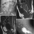
10.8.2 Scimitar Syndrome
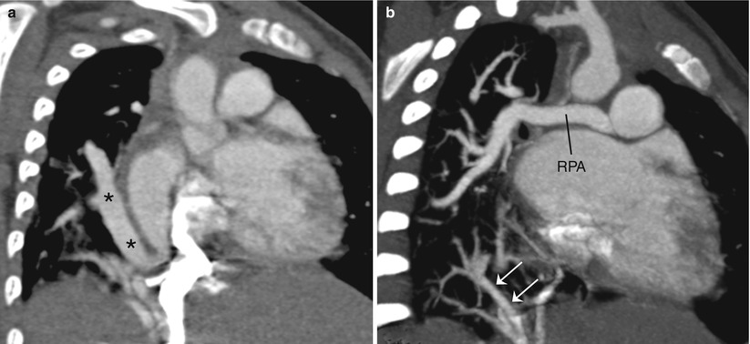
Fig. 10.2
(a, b) Scimitar syndrome. Coronal CT image demonstrates the scimitar vein (asterisks) anomalously draining into the right atrium–inferior vena cava junction (a). Coronal CT image shows the hypoplastic right pulmonary artery (RPA) and systemic arterial collaterals (arrows) supplying the right lower lung (b)
10.8.3 Horseshoe Lung
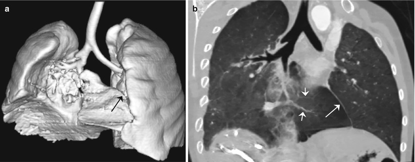
Fig. 10.3
(a, b) Horseshoe lung. Anterior shaded surface display CT image (a) and coronal minimum intensity projection CT image (b) show a fusion of both lungs and a pleural line (long arrow) between an isthmus of the hypoplastic right lung and the left lung. The pulmonary vasculature (short arrows) in the isthmic portion is derived from the hypoplastic lung (b)
10.8.4 Pulmonary Vein Atresia
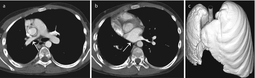
Fig. 10.4
(a–c) Right pulmonary vein atresia. Axial CT image at the pulmonary artery bifurcation level shows the hypoplastic right lung and right pulmonary artery (arrow) (a). Axial CT image at the left atrial level additionally reveals the absence of the right pulmonary vein (arrow) (b). Anterosuperior shaded surface display CT image demonstrates numerous reticular cracks on the hypoplastic right lung surface due to transpleural collateral arteries (c)
10.8.5 Congenital Diaphragmatic Hernia
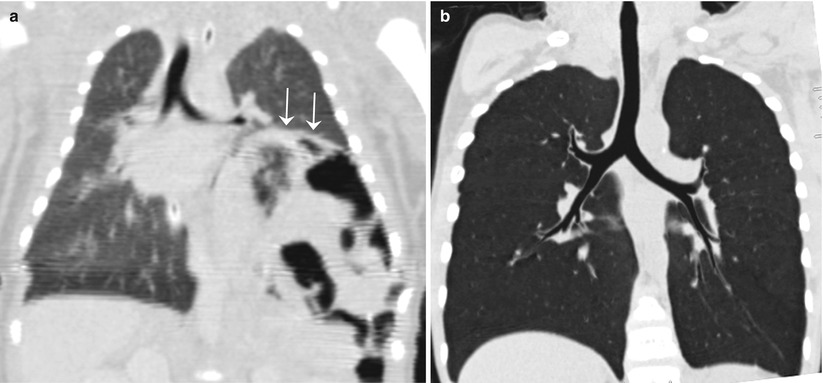
Fig. 10.5
(a, b) Congenital left diaphragmatic hernia. Coronal CT image shows the herniated abdominal contents (arrows) displacing the left lung upward (a). Coronal CT image 6 years after the diaphragmatic hernia repair demonstrates almost normal size of the left bronchi and compensatory hyperinflation of the left lung (b)
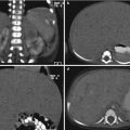
10.8.6 Bronchogenic Cyst
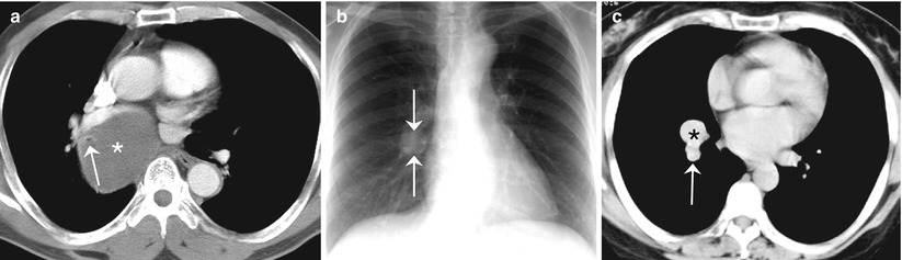
Fig. 10.6
(a–c) Bronchogenic cyst. Axial CT image shows a mediastinal cystic lesion (asterisk) encasing the right bronchus (arrow) and a mass effect on the adjacent structures (a). Plain chest radiograph reveals a well-circumscribed nodular lesion (arrows) in the medial portion of the right lower lung (b). Axial CT image demonstrates a hyperdense intrapulmonary cyst (asterisk) in the right lower lung anterior to the descending branch (arrow) of the right pulmonary artery (c)
10.8.7 Bronchial Atresia
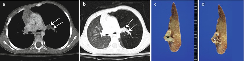
Fig. 10.7
(a–d) Bronchial atresia. Axial CT images with mediastinal (a) and lung (b) window settings show a hypodense mucocele (arrows) in the central area of the hyperinflated left upper lobe. Photographs show a resection specimen before (c) and after (d) the removal of mucus impacted in the dilated bronchus (asterisk)
10.8.8 Congenial Pulmonary Airway Malformation (CPAM)
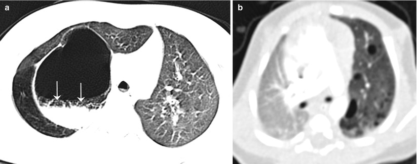
Fig. 10.8


(a, b) Congenial pulmonary airway malformation. Axial CT image shows a large thick-walled cyst with an air–fluid level (arrows) in the right lung that was proven as an infected congenital pulmonary airway malformation (type 1) (a). Axial CT image reveals type 2 congenital pulmonary airway malformation with multiple small cysts in the hyperlucent left lung (b)
Related posts:


Stay updated, free articles. Join our Telegram channel
Full access? Get Clinical Tree
