12.1 Cardiomegaly
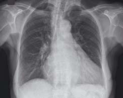
The heart is abnormally large. It takes up greater than 50% of the internal width of the thorax. The patient was known to have left ventricular failure
12.2 Pulmonary oedema
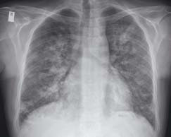
The heart is not enlarged but there are signs of pulmonary oedema with shadowing spreading from the hila. The patient was diabetic and presented with acute myocardial infarction and left ventricular failure
12.3 Kerley B lines
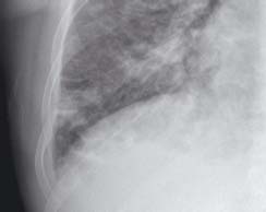
Close-up of costophrenic angle of fig 12.2. Here septal ‘Kerley B’ lines are seen. These are horizontal lines that reach the pleural surface. They are caused by fluid between the interlobular septa and are a specific sign of pulmonary oedema
12.4 Pleural effusions
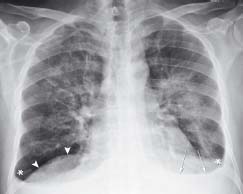
This patient with heart failure has pulmonary oedema and fluid is filling the pleural cavities forming pleural effusions (arrows). The costophrenic angles (*) are blunted. The dome of the diaphragm is still visible (arrowheads)
12.5 Prosthetic heart valves
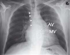
The heart is enlarged due to left ventricular failure. There is evidence of previous cardiac surgery with midline sternotomy wires (arrowheads) and metallic replacements of both the aortic valve (AV) and the mitral valve (MV)
12.6 Asbestos plaques
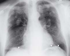
There are multiple dense irregular-shaped opacities over both lungs. These are typical appearances of asbestos plaques. The give-away sign is the layering of dense material over the diaphragm (arrowheads)
Cardiomegaly
Cardiomegaly is an abnormal enlargement of the heart where the maximum width of the heart shadow is greater than 50% of the maximum internal width of the thorax. The causes include:
- Hypertrophy – caused by an increase in afterload of a particular chamber (e.g. aortic stenosis, hypertension).
- Dilatation – secondary to toxic, metabolic, or infectious agents causing myocardial damage.
Stay updated, free articles. Join our Telegram channel

Full access? Get Clinical Tree


