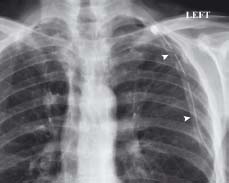
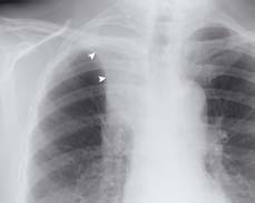
The edge of the left lung can be seen as a thin well-defined line (arrowheads). Beyond this line there are no further lung markings because air is collecting in the pleural cavity (*)
13.2 Chest drain
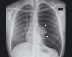
(Same patient as fig 13.1)
A tube has been inserted to drain air from the pleural cavity. It has been placed appropriately with its tip pointing high up towards the apex of the hemithorax
A tube has been inserted to drain air from the pleural cavity. It has been placed appropriately with its tip pointing high up towards the apex of the hemithorax
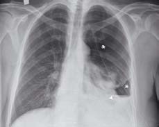
13.3 Tension pneumothorax
The left lung (arrowheads) is being compressed, the left hemidiaphragm is depressed, and the trachea (*) is pushed towards the right. Immediate aspiration is required
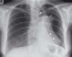
13.4 Hydropneumothorax
This patient has both a pneumothorax (*) and a pleural effusion (arrowhead) due to an oesophageal rupture. The fluid level does not have the curved meniscus sign of a simple effusion. Note the presence of a chest drain. Haemopneumothorax has identical appearances and is often associated with rib fractures
13.5 Lower left lobe collapse
The left lower lobe bronchus is occluded with collapse of this lobe. The edge of the collapsed lobe forms the ‘sail sign’ (arrowheads). The trachea (*) is pulled towards the side of volume loss
13.6 Right upper lobe collapse
The horizontal fissure (arrowheads) has been pulled upwards due to volume loss of the right upper lobe. The patients in both figs 13.5 and 13.6 had an occluding bronchial carcinoma
Pneumothorax
Pneumothorax is the presence of air in the pleural cavity. During normal ventilation, the thoracic volume increases to create relative negative intrathoracic pressure for lung inflation. However, in the presence of a pleural defect, air enters the potential pleural space and breaks this pressure potential, thereby compromising lung inflation. This impairs gaseous exchange and may cause breathlessness.
- Simple pneumothorax is common and occurs in healthy chests, especially in tall, slim, young males. Secondary pneumothoraces can arise in individuals with underlying disease, e.g. COPD, asthma, barotrauma, penetrating chest trauma and pneumonia.
- Tension pneumothorax
Related posts:

Stay updated, free articles. Join our Telegram channel


Full access? Get Clinical Tree


