Delayed-Enhancement Magnetic Resonance


  • Delayed-enhancement cardiac magnetic resonance (DE-CMR) can identify reversible myocardial dysfunction before coronary revascularization, predict improvement in contractile function in patients with reperfused acute myocardial infarction, and predict response to ß-blocker therapy in patients with heart failure.


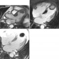
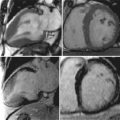
  • DE-CMR involves T1-weighted imaging of the heart after administration of gadolinium contrast media using a segmented inversion-recovery gradient echo sequence where infarcted or scarred myocardium accumulates gadolinium and appears as “hyperenhanced” or bright.


  • DE-CMR allows the identification of the presence, location, and extent of acute and chronic myocardial infarction with high accuracy relative to histopathology.


  • The pattern of hyperenhancement is useful in differentiating ischemic from nonischemic cardiomyopathy. Frequently the specific etiology responsible for nonischemic cardiomyopathy also may be ascertained.


  • DE-CMR improves the specificity and accuracy of stress perfusion CMR for the detection of coronary artery disease.


  • Emerging applications for DE-CMR include the detection of intracardiac thrombus and the assessment of patients with intraventricular dyssynchrony for potential cardiac resynchronization therapy.


  • Myocardial scarring detected by DE-CMR has been associated with adverse prognosis in patients both with ischemic and nonischemic heart disease.


KEY POINTS


Magnetic resonance imaging of the heart after administration of gadolinium contrast media has been described in the literature for over 20 years. This approach was predicated on the concept that injured tissue accumulates gadolinium and appears as “hyperenhanced” or bright on T1-weighted images acquired at least 10 minutes after injection of gadolinium. A major limitation of the initial approach was poor contrast between normal and injured myocardium. More recently a segmented inversion-recovery gradient echo sequence has been developed that significantly improves in vivo detection of hyperenhanced regions. This technique, known as delayed-enhancement cardiac magnetic resonance (DE-CMR), can identify the presence, location, and extent of both acute and chronic myocardial infarction with high spatial resolution. This technique has been extensively validated with comparisons to histopathology.


Accurate assessment of viable myocardium is important in patients with contractile dysfunction. In these patients if substantial viable myocardium is found, left ventricular function can improve following coronary revascularization and the functional improvement may be accompanied by survival benefits. Identification of nonviable myocardium is also important, because infarcted or scarred tissue may provide a substrate for ventricular tachyarrhythmias and sudden cardiac death. Thus the evaluation of both viable and nonviable myocardium provides important guidance for clinical decision making.


In this chapter we will first illustrate the physiologic basis for DE-CMR, and describe the protocol and typical imaging parameters. Then well-established applications of DE-CMR in patients with ischemic and nonischemic heart disease will be discussed, as well as some newer emerging applications. Finally recent reports demonstrating the prognostic significance of DE-CMR findings will be briefly reviewed.


TABLE 19-1

Typical DE-CMR Scanning Parameters


PARAMETER TYPICAL VALUES
Gadolinium dose 0.10-0.20 mmol/kg
Field of view 300-380 mm
In-plane voxel size 1.2-1.8 × 1.2-1.8 mm
Slice thickness 6 mm
Flip angle 20-30 degrees
Segments 13-31
Inversion time (IT) Variable
Bandwidth 90-250 Hz/pixel
Echo time (TE) 3-4 msec
Repetition time (TR) 8-9 msec
Gating factor 2
K-space ordering Linear
Fat saturation No
Asymmetric echo Yes
Gradient moment refocusing Yes

The dose of gadolinium given is usually 0.10 to 0.20 mmol/kg. Higher doses result in better SNR, but the bright LV blood pool may obscure subendocardial infarcts. The field-of-view (FOV) in both read and phase-encode directions is minimized to improve spatial resolution without resulting in wrap-around artifact in the area of interest. For patients with heart rates less than 90 beats per minute, we typically acquire 23 lines of k-space data during the mid-diastolic portion of the cardiac cycle. For a repetition time of 8 msec, the data acquisition window is 184 msec in duration (8 × 23 = 184). Because the middiastolic period of relative cardiac standstill is reduced in patients with faster heart rates, we decrease the number of segments (k-space lines) acquired per cardiac cycle in order to reduce the length of the imaging window. This eliminates blurring from cardiac motion during the k-space collection. In order to allow for adequate longitudinal relaxation between successive 180-degree inversion pulses, inversion pulses are applied every other heartbeat (gating factor of 2). In our experience an in-plane resolution of 1.2 to 1.8 mm by 1.2 to 1.8 mm with a slice thickness of 6 mm provides an adequate signal-to-noise balance while avoiding significant partial volume effects. As stated previously, the flip angle is kept shallow to retain the effects of the inversion prepulse, but it can be relatively greater (30 degrees) if larger doses of gadolinium are given (0.2 mmol/kg) and the IT of myocardium is correspondingly shorter.


Figure 19-1


The importance of myocardial viability assessment. This figure shows data pooled from 24 studies examining late survival with revascularization versus medical therapy after myocardial viability testing in patients with severe coronary artery disease and left ventricular dysfunction. This metaanalysis in 3088 patients demonstrated that in patients with viability, revascularization was associated with a 79.6% reduction in annual mortality (16% versus 3.2%, p less than 0.0001) compared with medical therapy. Patients without viability had intermediate mortality, trending to higher rates with revascularization versus medical therapy (7.7% versus 6.2%, p = NS). Mean left ventricular ejection fraction at baseline was 32 ± 8%, and patients were followed for 25 ± 10 months.

(From Allman et al: J Am Coll Cardiol 2002;39:1151-1158, with permission.)


Figure 19-2


Overall DE-CMR protocol. Imaging starts with scouting and typically a complete set of short- and long-axis cine images are acquired before contrast media is administered. Gadolinium is usually given at a dose of 0.1 to 0.2 mmol/kg of body weight. Ideally, delayed enhancement images are obtained at least 10 minutes after contrast administration in order to better separate bright myocardium from bright left ventricular blood pool. Cine and delayed enhancement images are taken at the same anatomic levels to enable direct comparison.


Figure 19-3


Typical viability scan. Interpretation is improved by alternating cine with delayed enhancement images to allow side-by-side comparison of dysfunctional regions (cine) with areas of infarction (delayed enhancement). In this patient example the diastolic still frames from the cine movies show regional thinning of the inferior and inferoseptal walls, which corresponds to the area of transmural infarction on the delayed enhancement images.


Figure 19-4


Comparison with histopathology. Multiple studies of both acute and chronic myocardial infarction have compared DE-CMR with histopathology and observed that the size and shape of hyperenhanced regions by DE-CMR closely match those of irreversible injury defined by histopathology. Short-axis histopathology sections of the heart are shown on the left, with corresponding delayed enhancement images on the right. The pale yellow/white regions on histopathology represent myocardial infarction, which closely match the bright areas on DE-CMR.

(From Fieno et al: J Am Coll Cardiol 2000;36:1985-1991, with permission.)


Figure 19-5


DE-CMR delineates irreversible from reversible injury. Three pathophysiologically distinct regions are depicted. Region “1” represents normal noninfarcted myocardium. Region “2” represents myocardium that has suffered temporary hypoperfusion and reversible injury—the “at risk but not infarcted” region. Viability of this region was confirmed by light microscopy. Region “3” represents infarcted myocardium. Panel A shows a portion of the left ventricular anterior wall following triphenyl tetrazolium chloride (TTC) staining for histopathology identification of viable myocardium. Panel B shows the same myocardial section under ultraviolet light. Note that both regions “2” and “3” lack fluorescent microparticles depicting hypoperfusion. Panel C shows ex vivo delayed enhancement imaging of the section. Importantly, region “2” does not hyperenhance by DE-CMR. This example demonstrates that areas suffering reversible injury do not hyperenhance by DE-CMR.

(From Fieno et al: J Am Coll Cardiol 2000;36:1985-1991, with permission.)


Figure 19-6


There is a close match between the extent of hyperenhancement by DE-CMR and infarct size ( blue circles ) in the acute (4 hours), subacute (10 days), and chronic (8 weeks) time points, independent of reperfusion status. Conversely, the area at risk ( red squares ) is nearly always larger than the area of hyperenhancement. In this study the infarct size was determined by triphenyl tetrazolium chloride (TTC) staining and the myocardium at risk of infarction was identified by injecting fluorescent microparticles into the left atrium after reoccluding the coronary artery before sacrificing the animal for histopathology. These data are consistent with the principle that hyperenhancement only occurs in regions of irreversible injury.

(Adapted from Fieno et al: J Am Coll Cardiol 2000;36:1985-1991, with permission.)


Figure 19-7


The physiologic basis of hyperenhancement in DE-CMR. In normal myocardium, two points should be noted. First, myocytes are densely packed, and total tissue volume is predominantly intracellular (≈ 80% of the water space). Second, gadolinium contrast media does not cross cellular membranes, and thus is limited to the extracellular (intravascular and interstitial) space. The result is that the volume of distribution of gadolinium contrast in normal myocardium is quite small (≈ 20% of water space), and one can consider viable myocytes as actively excluding contrast media. In acutely infarcted regions the myocyte membranes are ruptured, allowing gadolinium to passively diffuse into the intracellular space. The result is an increased concentration of gadolinium at the tissue level and therefore hyperenhancement occurs. Myocardial scar is characterized by a dense collagenous matrix; however, at a cellular level the interstitial space between collagen fibers may be significantly greater than the interstitial space between the living myocytes that is characteristic of normal myocardium. Thus the concentration of gadolinium in scar is greater than in normal myocardium because of the expanded volume of distribution and the regions of scar appear hyperenhanced by DE-CMR.


Figure 19-8


Comparison of single photon emission computed tomography (SPECT) and DE-CMR with histology in three canines with subendocardial infarcts ( arrows ). Note that the infarcts are not evident on SPECT. It is often reported that the mechanism by which subendocardial infarcts are routinely missed by SPECT is limited spatial resolution. However, the primary mechanism may relate to the inability of SPECT to directly visualize both viable and infarcted myocardium. Measuring only the amount of viability is problematic because there is a large intrinsic variation in the amount of viability in normal regions. Hence, although tracer activity “on average” may be modestly reduced in the setting of subendocardial infarction, for any specific patient or region, the level of reduction may be smaller than the normal regional variation, rendering the subendocardial infarction “invisible.” With DE-CMR, both viable (unenhanced) and infarcted (enhanced) myocardium are visualized simultaneously. Note on the DE-CMR images that hyperenhanced regions correspond to infarcted regions by histopathology. Note also, on both DE-CMR and histopathology, that even the regions completely free of infarction have variable wall thickness, representing variable absolute amounts of viability.

(Adapted from Wagner et al: Lancet 2003;361:374-379, with permission.)


Feb 1, 2019 | Posted by in MAGNETIC RESONANCE IMAGING | Comments Off on Delayed-Enhancement Magnetic Resonance

Full access? Get Clinical Tree

Get Clinical Tree app for offline access