Chapter 13
Desmoid Fibromatosis
Epidemiology
Desmoid fibromatosis is arbitrarily divided into two types: anterior abdominal wall and extra-abdominal wall. The extra-abdominal variety, which is more common, is also known as musculoaponeurotic or aggressive fibromatosis. The histologic appearances of these varieties are the same. Approximately 10 to 30% of all extra-abdominal desmoids are found in the head and neck. Desmoid fibromatosis affects a wide age group ranging from infants to the eighth decade. Most patients, however, are in the third and fourth decades. There is no sex predilection for the extra-abdominal variety although there is a female preponderance in the abdominal type.
Clinical Findings
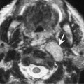
The lesion is firm to hard and is characteristically slow growing. Most lesions are nontender and painless. They may be noted to develop in previously irradiated fields or surgical scars. Some lesions are multicentric. Lesions resemble scar and may be impossible to distinguish from proliferating scar tissue both clinically and pathologically.
Pathology
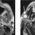
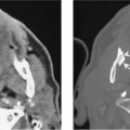
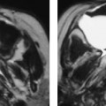
The lesions are variable in size and may grow beyond 20 cm. They develop within muscles, aponeurosis, or fascia and typically infiltrate the muscles along the long axis. Microscopically, the muscles and aponeurosis are invaded by mature, uniform, spindle-shaped cells. The infiltrative process separates the muscle bundles and these muscles eventually show atrophy. In some patients these lesions turn sarcomatous.
Treatment