In up to 40% of ischemic stroke cases the etiology remains unknown. A substantial proportion of these patients has non- or only mildly stenosing carotid artery plaques not fulfilling common criteria for large artery stroke, but beeing suspicious for arterio-arteriell embolism. Several imaging techniques allow the non-invasive analysis of plaque features. Nevertheless, carotid MRI might be best suited to assess the key features of vulnerable plaques. This review article discusses potential causes of cryptogenic stroke, the role of plaque imaging in non-stenosing plaques and the association of vulnerable plaques and specific plaque features with stroke risk and stroke recurrence.
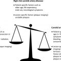
In patients with mild and nonstenosing plaques, high-resolution carotid MR imaging might be the most promising tool to assess the correlation of vulnerable plaques and cryptogenic stroke, stroke recurrence, and plaque progression.
In patients with mild and nonstenosing plaques, high-resolution carotid MR imaging might be the most promising tool to assess the correlation of vulnerable plaques and cryptogenic stroke, stroke recurrence, and plaque progression.
References
- 1. Rosamond W., Flegal K., Furie K., et al: Heart disease and stroke statistics–2008 update: a report from the American Heart Association Statistics Committee and Stroke Statistics Subcommittee. Circulation 2008; 117: pp. e25-146
- 2. Brott T.G., Halperin J.L., Abbara S., et al: 2011 ASA/ACCF/AHA/AANN/AANS/ACR/ASNR/CNS/SAIP/SCAI/SIR/SNIS/SVM/SVS guideline on the management of patients with extracranial carotid and vertebral artery disease: executive summary: a report of the American College of Cardiology Foundation/American Heart Association Task Force on Practice Guidelines, and the American Stroke Association, American Association of Neuroscience Nurses, American Association of Neurological Surgeons, American College of Radiology, American Society of Neuroradiology, Congress of Neurological Surgeons, Society of Atherosclerosis Imaging and Prevention, Society for Cardiovascular Angiography and Interventions, Society of Interventional Radiology, Society of NeuroInterventional Surgery, Society for Vascular Medicine, and Society for Vascular Surgery. J Am Coll Cardiol 2011; 57: pp. 1002-1044
- 3. Falk E., Shah P.K., and Fuster V.: Coronary plaque disruption. Circulation 1995; 92: pp. 657-671
- 4. Spagnoli L.G., Mauriello A., Sangiorgi G., et al: Extracranial thrombotically active carotid plaque as a risk factor for ischemic stroke. JAMA 2004; 292: pp. 1845-1852
- 5. Putaala J., Curtze S., Hiltunen S., et al: Causes of death and predictors of 5-year mortality in young adults after first-ever ischemic stroke: the Helsinki Young Stroke Registry. Stroke 2009; 40: pp. 2698-2703
- 6. Adams H.P., Bendixen B.H., Kappelle L.J., et al: Classification of subtype of acute ischemic stroke. Definitions for use in a multicenter clinical trial. TOAST. Trial of Org 10172 in Acute Stroke Treatment. Stroke 1993; 24: pp. 35-41
- 7. Amarenco P., Bogousslavsky J., Caplan L.R., et al: New approach to stroke subtyping: the A-S-C-O (phenotypic) classification of stroke. Cerebrovasc Dis 2009; 27: pp. 502-508
- 8. Eriksson S.E., and Olsson J.E.: Survival and recurrent strokes in patients with different subtypes of stroke: a fourteen-year follow-up study. Cerebrovasc Dis 2001; 12: pp. 171-180
- 9. Bang O.Y., Lee P.H., Joo S.Y., et al: Frequency and mechanisms of stroke recurrence after cryptogenic stroke. Ann Neurol 2003; 54: pp. 227-234
- 10. Yaghi S., and Elkind M.S.: Cryptogenic stroke: a diagnostic challenge. Neurol Clin Pract 2014; 4: pp. 386-393
- 11. Hart R.G., Diener H.C., Coutts S.B., et al: Embolic strokes of undetermined source: the case for a new clinical construct. Lancet Neurol 2014; 13: pp. 429-438
- 12. Ruff C.T., Giugliano R.P., Braunwald E., et al: Comparison of the efficacy and safety of new oral anticoagulants with warfarin in patients with atrial fibrillation: a meta-analysis of randomised trials. Lancet 2014; 383: pp. 955-962
- 13. Sanna T., Diener H.C., Passman R.S., et al: Cryptogenic stroke and underlying atrial fibrillation. N Engl J Med 2014; 370: pp. 2478-2486
- 14. Rizos T., Guntner J., Jenetzky E., et al: Continuous stroke unit electrocardiographic monitoring versus 24-hour Holter electrocardiography for detection of paroxysmal atrial fibrillation after stroke. Stroke 2012; 43: pp. 2689-2694
- 15. Handke M., Harloff A., Olschewski M., et al: Patent foramen ovale and cryptogenic stroke in older patients. N Engl J Med 2007; 357: pp. 2262-2268
- 16. Di Tullio M.R., Sacco R.L., Sciacca R.R., et al: Patent foramen ovale and the risk of ischemic stroke in a multiethnic population. J Am Coll Cardiol 2007; 49: pp. 797-802
- 17. Kronzon I., and Tunick P.A.: Aortic atherosclerotic disease and stroke. Circulation 2006; 114: pp. 63-75
- 18. Wehrum T., Kams M., Strecker C., et al: Prevalence of potential retrograde embolization pathways in the proximal descending aorta in stroke patients and controls. Cerebrovasc Dis 2014; 38: pp. 410-417
- 19. Geroulakos G., Ramaswami G., Nicolaides A., et al: Characterization of symptomatic and asymptomatic carotid plaques using high-resolution real-time ultrasonography. Br J Surg 1993; 80: pp. 1274-1277
- 20. Sztajzel R., Momjian S., Momjian-Mayor I., et al: Stratified gray-scale median analysis and color mapping of the carotid plaque: correlation with endarterectomy specimen histology of 28 patients. Stroke 2005; 36: pp. 741-745
- 21. Russell D.A., Wijeyaratne S.M., and Gough M.J.: Relationship of carotid plaque echomorphology to presenting symptom. Eur J Vasc Endovasc Surg 2010; 39: pp. 134-138
- 22. Topakian R., King A., Kwon S.U., et al: Ultrasonic plaque echolucency and emboli signals predict stroke in asymptomatic carotid stenosis. Neurology 2011; 77: pp. 751-758
- 23. Hallerstam S., Carlstrom C., Zetterling M., et al: Carotid atherosclerosis in relation to symptoms from the territory supplied by the carotid artery. Eur J Vasc Endovasc Surg 2000; 19: pp. 356-361
- 24. Reiter M., Effenberger I., Sabeti S., et al: Increasing carotid plaque echolucency is predictive of cardiovascular events in high-risk patients. Radiology 2008; 248: pp. 1050-1055
- 25. Kakkos S.K., Nicolaides A.N., Kyriacou E., et al: Computerized texture analysis of carotid plaque ultrasonic images can identify unstable plaques associated with ipsilateral neurological symptoms. Angiology 2011; 62: pp. 317-328
- 26. Griffin M.B., Kyriacou E., Pattichis C., et al: Juxtaluminal hypoechoic area in ultrasonic images of carotid plaques and hemispheric symptoms. J Vasc Surg 2010; 52: pp. 69-76
- 27. Ainsworth C.D., Blake C.C., Tamayo A., et al: 3D ultrasound measurement of change in carotid plaque volume: a tool for rapid evaluation of new therapies. Stroke 2005; 36: pp. 1904-1909
- 28. AlMuhanna K., Hossain M.M., Zhao L., et al: Carotid plaque morphometric assessment with three-dimensional ultrasound imaging. J Vasc Surg 2015; 61: pp. 690-697
- 29. Markus H.S., and MacKinnon A.: Asymptomatic embolization detected by Doppler ultrasound predicts stroke risk in symptomatic carotid artery stenosis. Stroke 2005; 36: pp. 971-975
- 30. Markus H.S., Droste D.W., Kaps M., et al: Dual antiplatelet therapy with clopidogrel and aspirin in symptomatic carotid stenosis evaluated using Doppler embolic signal detection: the Clopidogrel and Aspirin for Reduction of Emboli in Symptomatic Carotid Stenosis (CARESS) trial. Circulation 2005; 111: pp. 2233-2240
- 31. Ritter M.A., Dittrich R., Thoenissen N., et al: Prevalence and prognostic impact of microembolic signals in arterial sources of embolism. A systematic review of the literature. J Neurol 2008; 255: pp. 953-961
- 32. Shalhoub J., Owen D.R., Gauthier T., et al: The use of contrast enhanced ultrasound in carotid arterial disease. Eur J Vasc Endovasc Surg 2010; 39: pp. 381-387
- 33. Shah F., Balan P., Weinberg M., et al: Contrast-enhanced ultrasound imaging of atherosclerotic carotid plaque neovascularization: a new surrogate marker of atherosclerosis? Vasc Med 2007; 12: pp. 291-297
- 34. Li C., He W., Guo D., et al: Quantification of carotid plaque neovascularization using contrast-enhanced ultrasound with histopathologic validation. Ultrasound Med Biol 2014; 40: pp. 1827-1833
- 35. Hoogi A., Adam D., Hoffman A., et al: Carotid plaque vulnerability: quantification of neovascularization on contrast-enhanced ultrasound with histopathologic correlation. AJR Am J Roentgenol 2011; 196: pp. 431-436
- 36. Xiong L., Deng Y.B., Zhu Y., et al: Correlation of carotid plaque neovascularization detected by using contrast-enhanced US with clinical symptoms. Radiology 2009; 251: pp. 583-589
- 37. Ritter M.A., Theismann K., Schmiedel M., et al: Vascularization of carotid plaque in recently symptomatic patients is associated with the occurrence of transcranial microembolic signals. Eur J Neurol 2013; 20: pp. 1218-1221
- 38. Shalhoub J., Monaco C., Owen D.R., et al: Late-phase contrast-enhanced ultrasound reflects biological features of instability in human carotid atherosclerosis. Stroke 2011; 42: pp. 3634-3636
- 39. Owen D.R., Shalhoub J., Miller S., et al: Inflammation within carotid atherosclerotic plaque: assessment with late-phase contrast-enhanced US. Radiology 2010; 255: pp. 638-644
- 40. Serfaty J.M., Nonent M., Nighoghossian N., et al: Plaque density on CT, a potential marker of ischemic stroke. Neurology 2006; 66: pp. 118-120
- 41. Trelles M., Eberhardt K.M., Buchholz M., et al: CTA for screening of complicated atherosclerotic carotid plaque–American Heart Association type VI lesions as defined by MRI. AJNR Am J Neuroradiol 2013; 34: pp. 2331-2337
- 42. Gupta A., Baradaran H., Kamel H., et al: Evaluation of computed tomography angiography plaque thickness measurements in high-grade carotid artery stenosis. Stroke 2014; 45: pp. 740-745
- 43. Gupta A., Mtui E.E., Baradaran H., et al: CT angiographic features of symptom-producing plaque in moderate-grade carotid artery stenosis. AJNR Am J Neuroradiol 2015; 36: pp. 349-354
- 44. Gupta A., Baradaran H., Mtui E.E., et al: Detection of symptomatic carotid plaque using source data from MR and CT angiography: a correlative study. Cerebrovasc Dis 2015; 39: pp. 151-161
- 45. Saba L., and Mallarini G.: Carotid plaque enhancement and symptom correlations: an evaluation by using multidetector row CT angiography. AJNR Am J Neuroradiol 2011; 32: pp. 1919-1925
- 46. Saba L., Piga M., Raz E., et al: Carotid artery plaque classification: does contrast enhancement play a significant role? AJNR Am J Neuroradiol 2012; 33: pp. 1814-1817
- 47. Rudd J.H., Warburton E.A., Fryer T.D., et al: Imaging atherosclerotic plaque inflammation with [18F]-fluorodeoxyglucose positron emission tomography. Circulation 2002; 105: pp. 2708-2711
- 48. Judenhofer M.S., Wehrl H.F., Newport D.F., et al: Simultaneous PET-MRI: a new approach for functional and morphological imaging. Nat Med 2008; 14: pp. 459-465
- 49. Marnane M., Merwick A., Sheehan O.C., et al: Carotid plaque inflammation on 18F-fluorodeoxyglucose positron emission tomography predicts early stroke recurrence. Ann Neurol 2012; 71: pp. 709-718
- 50. Müller H.F., Viaccoz A., Fisch L., et al: 18FDG-PET-CT: an imaging biomarker of high-risk carotid plaques. Correlation to symptoms and microembolic signals. Stroke 2014; 45: pp. 3561-3566
- 51. Truijman M.T., Kwee R.M., van Hoof R.H., et al: Combined 18F-FDG PET-CT and DCE-MRI to assess inflammation and microvascularization in atherosclerotic plaques. Stroke 2013; 44: pp. 3568-3570
- 52. Yuan C., Zhang S.X., Polissar N.L., et al: Identification of fibrous cap rupture with magnetic resonance imaging is highly associated with recent transient ischemic attack or stroke. Circulation 2002; 105: pp. 181-185
- 53. Cai J.M., Hatsukami T.S., Ferguson M.S., et al: Classification of human carotid atherosclerotic lesions with in vivo multicontrast magnetic resonance imaging. Circulation 2002; 106: pp. 1368-1373
- 54. Takaya N., Yuan C., Chu B., et al: Association between carotid plaque characteristics and subsequent ischemic cerebrovascular events: a prospective assessment with MRI–initial results. Stroke 2006; 37: pp. 818-823
- 55. Saam T., Underhill H.R., Chu B., et al: Prevalence of American Heart Association type VI carotid atherosclerotic lesions identified by magnetic resonance imaging for different levels of stenosis as measured by duplex ultrasound. J Am Coll Cardiol 2008; 51: pp. 1014-1021
- 56. Saam T., Hetterich H., Hoffmann V., et al: Meta-analysis and systematic review of the predictive value of carotid plaque hemorrhage on cerebrovascular events by magnetic resonance imaging. J Am Coll Cardiol 2013; 62: pp. 1081-1091
- 57. Parmar J.P., Rogers W.J., Mugler J.P., et al: Magnetic resonance imaging of carotid atherosclerotic plaque in clinically suspected acute transient ischemic attack and acute ischemic stroke. Circulation 2010; 122: pp. 2031-2038
- 58. Zhao H., Zhao X., Liu X., et al: Association of carotid atherosclerotic plaque features with acute ischemic stroke: a magnetic resonance imaging study. Eur J Radiol 2013; 82: pp. e465-70
- 59. Freilinger T.M., Schindler A., Schmidt C., et al: Prevalence of nonstenosing, complicated atherosclerotic plaques in cryptogenic stroke. JACC Cardiovasc Imaging 2012; 5: pp. 397-405
- 60. Lindsay A.C., Biasiolli L., Lee J.M., et al: Plaque features associated with increased cerebral infarction after minor stroke and TIA: a prospective, case-control, 3-T carotid artery MR imaging study. JACC Cardiovasc Imaging 2012; 5: pp. 388-396
- 61. McNally J.S., McLaughlin M.S., Hinckley P.J., et al: Intraluminal thrombus, intraplaque hemorrhage, plaque thickness, and current smoking optimally predict carotid stroke. Stroke 2015; 46: pp. 84-90
- 62. Kwee R.M., van Oostenbrugge R.J., Prins M.H., et al: Symptomatic patients with mild and moderate carotid stenosis: plaque features at MRI and association with cardiovascular risk factors and statin use. Stroke 2010; 41: pp. 1389-1393
- 63. Cheung H.M., Moody A.R., Singh N., et al: Late stage complicated atheroma in low-grade stenotic carotid disease: MR imaging depiction–prevalence and risk factors. Radiology 2011; 260: pp. 841-847
- 64. Singh N., Moody A.R., Gladstone D.J., et al: Moderate carotid artery stenosis: MR imaging-depicted intraplaque hemorrhage predicts risk of cerebrovascular ischemic events in asymptomatic men. Radiology 2009; 252: pp. 502-508
- 65. Kolodgie F.D., Gold H.K., Burke A.P., et al: Intraplaque hemorrhage and progression of coronary atheroma. N Engl J Med 2003; 349: pp. 2316-2325
- 66. Takaya N., Yuan C., Chu B., et al: Presence of intraplaque hemorrhage stimulates progression of carotid atherosclerotic plaques: a high-resolution magnetic resonance imaging study. Circulation 2005; 111: pp. 2768-2775
- 67. Underhill H.R., Yuan C., Yarnykh V.L., et al: Arterial remodeling in [corrected] subclinical carotid artery disease. JACC Cardiovasc Imaging 2009; 2: pp. 1381-1389
- 68. Kwee R.M., van Oostenbrugge R.J., Mess W.H., et al: Carotid plaques in transient ischemic attack and stroke patients: one-year follow-up study by magnetic resonance imaging. Invest Radiol 2010; 45: pp. 803-809
- 69. Saam T., Yuan C., Chu B., et al: Predictors of carotid atherosclerotic plaque progression as measured by noninvasive magnetic resonance imaging. Atherosclerosis 2007; 194: pp. e34-42
- 70. Migrino R.Q., Bowers M., Harmann L., et al: Carotid plaque regression following 6-month statin therapy assessed by 3T cardiovascular magnetic resonance: comparison with ultrasound intima media thickness. J Cardiovasc Magn Reson 2011; 13: pp. 37
- 71. Boussel L., Arora S., Rapp J., et al: Atherosclerotic plaque progression in carotid arteries: monitoring with high-spatial-resolution MR imaging–multicenter trial. Radiology 2009; 252: pp. 789-796
- 72. Zhao X.Q., Dong L., Hatsukami T., et al: MR imaging of carotid plaque composition during lipid-lowering therapy a prospective assessment of effect and time course. JACC Cardiovasc Imaging 2011; 4: pp. 977-986
- 73. Underhill H.R., Yuan C., Zhao X.Q., et al: Effect of rosuvastatin therapy on carotid plaque morphology and composition in moderately hypercholesterolemic patients: a high-resolution magnetic resonance imaging trial. Am Heart J 2008; 155: pp. 584.e1-584.e8
- 74. Yamaguchi Oura M., Sasaki M., Ohba H., et al: Carotid plaque characteristics on magnetic resonance plaque imaging following long-term cilostazol therapy. J Stroke Cerebrovasc Dis 2014; 23: pp. 2425-2430
- 75. Altaf N., Daniels L., Morgan P.S., et al: Detection of intraplaque hemorrhage by magnetic resonance imaging in symptomatic patients with mild to moderate carotid stenosis predicts recurrent neurological events. J Vasc Surg 2008; 47: pp. 337-342
- 76. Teng Z., Sadat U., Huang Y., et al: In vivo MRI-based 3D mechanical stress-strain profiles of carotid plaques with juxtaluminal plaque haemorrhage: an exploratory study for the mechanism of subsequent cerebrovascular events. Eur J Vasc Endovasc Surg 2011; 42: pp. 427-433
- 77. Teng Z., Degnan A.J., Sadat U., et al: Characterization of healing following atherosclerotic carotid plaque rupture in acutely symptomatic patients: an exploratory study using in vivo cardiovascular magnetic resonance. J Cardiovasc Magn Reson 2011; 13: pp. 64
- 78. Truijman M.T., Kooi M.E., van Dijk A.C., et al: Plaque At RISK (PARISK): prospective multicenter study to improve diagnosis of high-risk carotid plaques. Int J Stroke 2014; 9: pp. 747-754
- 79. Truijman M.T., de Rotte A.A., Aaslid R., et al: Intraplaque hemorrhage, fibrous cap status, and microembolic signals in symptomatic patients with mild to moderate carotid artery stenosis: the Plaque at RISK study. Stroke 2014; 45: pp. 3423-3426
- 80. Bayer-Karpinska A., Schwarz F., Wollenweber F.A., et al: The carotid plaque imaging in acute stroke (CAPIAS) study: protocol and initial baseline data. BMC Neurol 2013; 13: pp. 201
- 81. Schwarz F., Bayer-Karpinska A., Poppert H., et al: Serial carotid MRI identifies rupture of a vulnerable plaque resulting in amaurosis fugax. Neurology 2013; 80: pp. 1171-1172
Related posts:
 Three-Dimensional Carotid Plaque MR Imaging
Incorporating Carotid Plaque Imaging into Routine Clinical Carotid Magnetic Resonance Angiography
FDG PET/CT Imaging of Carotid Atherosclerosis
Clinical Perspective of Carotid Plaque Imaging
Three-Dimensional Carotid Plaque MR Imaging
Incorporating Carotid Plaque Imaging into Routine Clinical Carotid Magnetic Resonance Angiography
FDG PET/CT Imaging of Carotid Atherosclerosis
Clinical Perspective of Carotid Plaque Imaging
 Plaque Imaging to Decide on Optimal Treatment
 Low-Grade Carotid Stenosis
Stay updated, free articles. Join our Telegram channel

Full access? Get Clinical Tree


