Development
Mammals (from the Latin Mammalia) are named for the fact that they feed their young on milk produced in the mammary gland. The various species of mammals have developed different numbers of mammary glands along the genetically determined milk line.
Mammalian breasts generally occur in pairs, as is the case in humans. Normally, humans have one breast on each side of the chest. At the prepubescent stage, both girls and boys have mammary buds. With the onset of puberty, the production of estrogen increases in the female body, causing changes in the mammary gland: the breast buds enlarge and the breast exhibits an increased build-up of adipose tissue. Milk is secreted in the mammary gland even when there is no pregnancy, but the volume of this basic secretion varies widely from one individual to the next, and often goes unnoticed.
1.2 Anatomy
The network of lactiferous ducts resembles a coral bush (▶ Fig. 1.1). From the point of exit at the nipple (mammilla), the milk ducts branch out within the breast, with a smaller duct diameter after each divergence. At the periphery, the smallest milk ducts originate from the lobules of the gland, where the milk is secreted. Some 30 of these milk ducts, leading from all four quadrants of the breast, open out into the mammilla and thus form the functional unit of the mammary gland.
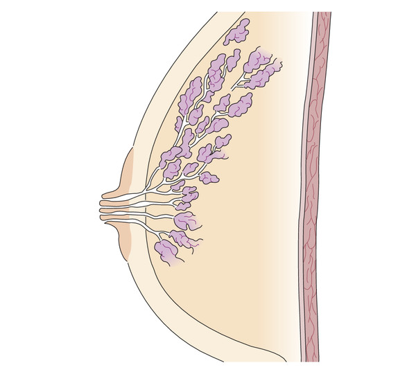
Fig. 1.1 The course of a milk duct. A mammary duct divides numerous times from the nipple. The smallest milk ducts originate from the lobules of the gland at the periphery and the milk is conducted from many lobules to the mammilla.
Stay updated, free articles. Join our Telegram channel

Full access? Get Clinical Tree


