(1)
Department of Pediatric Radiology, University of Texas Medical Branch, Galveston, TX, USA
Abstract
This chapter deals with developmental anatomy of the cervical spine. It emphasizes normal structures that are seen in the growing spine, which often can be misinterpreted for pathology.
The third through seventh vertebrae of the cervical spine are similar in their development, but development is different for the first and second vertebrae. All the cervical vertebrae develop from primitive sclerotomes [1], but the upper cervical spine, because of its functional complexity evolves from both the occipital and upper cervical sclerotomes (Fig. 1.1). These sclerotomes undergo significant embryologic alterations so as to eventually be able to accommodate free movement of the head on the first two (atlas and axis) cervical vertebrae. In this regard, the skull first must sit firmly on the atlas. This is accomplished by way of the occipital condyles articulating with the lateral masses of C1. This union is ensured by the presence of strong ligaments between these two structures. Thereafter, the skull and C1 must be able to rotate freely on C2. This is facilitated by the dens, which serves as a pivot for this function. The resultant rotatory movement occurs primarily at the C1–C2 level, but it should be underscored that while C1 rotates, within limits, around the dens, on flexion and extension, the skull, C1, and C2 move as a unit.
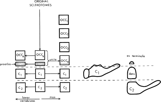

Fig. 1.1
Embryologic development of the upper cervical spine. The skull and upper cervical spine develop from primitive sclerotomes; the skull from occipital sclerotomes and the spine from upper cervical sclertomes. The occipital sclerotomes tend to fuse and reduce in number which often is accompanied by deletion of some of their more primordial elements. It is important to note, however, that the primitive proatlas prevails and unites with the primordial fourth occipital sclerotome to form the final occipital sclerotome. The os terminale is derived from this sclerotome. The first vertebral body (C1) is derived from the first cervical sclerotome. The dens actually is the body of C1, but eventually it unites with the body of C2, which is derived from the second cervical sclerotome
In terms of the derivation of the various portions of the upper cervical spine from the primitive sclerotomes, the atlas (C1) is derived from the first cervical sclerotome (Fig. 1.1). The axis (C2) is derived from the second cervical sclerotome. This sclerotome gives rise to the body, lateral masses, and neural arch of C2, but not the dens. The dens actually is the body of C1 and as such is derived from the first cervical sclerotome. The ossicle at the tip of the dens (the os terminale) is derived from the fourth occipital sclerotome (specifically known as the proatlas). The dens itself arises from two primordial centers, which eventually fuse with the os terminale to form the mature dens (Fig. 1.2). If they remain separate the dens often is referred to as bifid, but still normal (see Fig. 4.19)
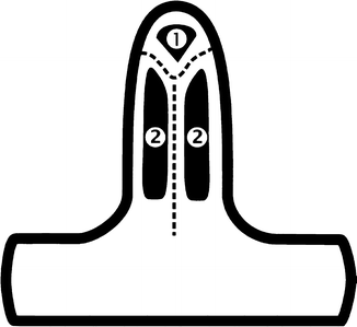

Fig. 1.2
Ossification centers of the dens. Note the two centers (2) for the body of the dens and the single center (1) for the tip, or os terminale
In terms of the development of the third through seventh cervical vertebra, and actually the body and neural arch of C2, there are six chondrofication centers [2]. These are diagrammatically depicted in Fig. 1.3, and while in most cases all these centers unite, if they fail to unite or develop, predictable anomalous configurations result. For example, if the two vertebral body centers fail to completely unite, a sagittal cleft vertebra results (Fig. 1.3b). If the union of the two vertebral body chondrofication centers is more advanced, but yet incomplete, a so-called butterfly vertebra develops (see Fig. 3.1), and when one or other, or both, of the vertebral body chondrofication centers fail to develop, a hemivertebra, or absence of a vertebra, results (Fig. 1.3c, d). Failure of development of the midchondrofication center leads to the absence of the pedicle (Fig. 1.3e), and if failure of the chondrofication center of the neural arch occurs, partial or complete absence of the neural arch results (Fig. 1.3f, g). Finally, if the two neural arches fail to unite posteriorly, a spina bifida results (Fig. 1.3h). However, development of the vertebral bodies at this stage is not complete for later, two vertebral body ossification centers develop. One is ventral and the other dorsal (Fig. 1.4a). If the ossification centers develop but fail to fuse, a coronal cleft vertebra results (Fig. 1.4b). If one or other of these ossification centers fails to develop, a coronal hemivertebra results (Fig. 1.4c, d). If both ossification centers fail to develop, the vertebral body will be hypoplastic or absent. Failure of the ossification centers of the neural arches to develop results in anomalies similar to those seen when the corresponding chondrofication centers fail to develop.
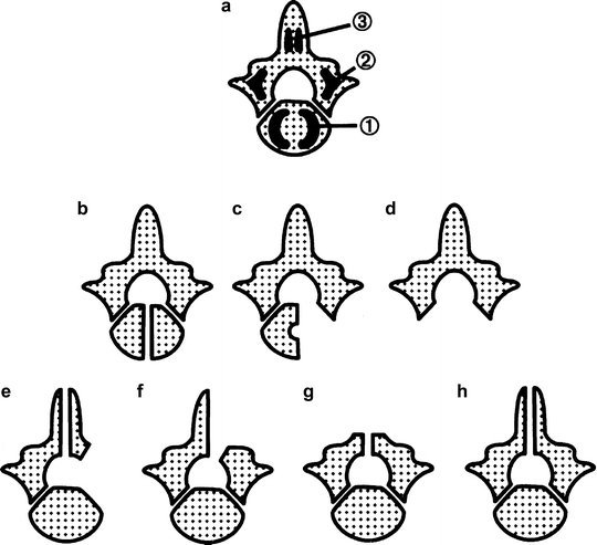


Fig. 1.3
Embryologic development of the cervical spine, chondrofication stage. (a) Note three chondrofication centers on each side: body (1), pars (2), and neural arch (3). (b) Lack of union of the body centers results in a sagittal cleft (complete), or butterfly (incomplete) vertebra. (c) Sagittal hemivertebra resulting from failure of development of one of the vertebral body centers. (d) Absent vertebra. Failure of development of both body centers. (e) Defect of pars. The center for the pars interarticularis fails to develop. (f) Lack of one center for the posterior arch usually results in half of the neural arch being present. (g) Absence of both posterior arch centers resulting in absence of the posterior arch. (h) Incomplete union of posterior arch (spina bifida)
< div class='tao-gold-member'>
Only gold members can continue reading. Log In or Register to continue
Stay updated, free articles. Join our Telegram channel

Full access? Get Clinical Tree


