Diagnostic Coronary Angiography

CHAPTER 7 Diagnostic Coronary Angiography


Despite significant advances in coronary CT angiography and MR angiography in the past decade, coronary angiography is the mainstay for the evaluation of coronary artery morphology and disease, and is a mandatory prerequisite for coronary intervention. Therefore, it is useful to have an appreciation for its strengths and limitations relative to coronary CT angiography.


TECHNICAL REQUIREMENTS


High-quality coronary angiography requires excellent quality fluoroscopic and digital imaging and a C-arm capable of three-dimensional rotation. These may be either single plane or biplane. Biplane laboratories are most useful for pediatric angiography and studies where simultaneous biplane fluoroscopy is needed (e.g., electrophysiologic studies). The x-ray tube must have a high heat capacity and small and large focal spots so that large patients can be imaged. Digital image receptors should have multiple magnification modes so that the entire heart can be imaged with a large field of view, and the coronary arteries can be imaged with a smaller field of view at higher spatial resolution. The use of higher magnification modes requires more panning (table movement during angiography) and higher radiation dose. As in static image systems, a grid is placed on the face of the image intensifier to reduce the effect of scatter radiation on the radiographic image. Coronary cineangiograms are typically recorded at 15 frames/sec, 30 frames/sec, or 60 frames/sec depending on the indication for the study.


In the past, images were recorded on 35-mm cine film and viewed on a cine projector. Today, catheterization laboratories use digital recording with viewing on a computer monitor. Although the spatial resolution of cine film is higher, digital recording is so much more convenient that it has become the standard. Digital recording does not require film processing with its inherent requirements for quality control, and the storage space required for cine film and the cost of cine film are enormous compared with digital media storage and cost.


ANGIOGRAPHIC VIEWS


Various angiographic projections are used to achieve the goal of viewing each coronary segment in at least two projections, free of foreshortening and free of overlap with other vessels. This goal was impossible before the advent of C-arms capable of triaxial motion. With modern equipment, angiographic views are limited only by physical impingement of the x-ray tube or image receptor on the patient or table, and the patient’s body habitus. In addition to rotation in the axial plane (generally from about 45 degrees right anterior oblique [RAO] to left lateral), the image receptor must be positioned over the patient’s shoulder to produce cranially angulated views and over the hip to produce caudally angulated views to image many coronary artery segments adequately. Cranial or caudal angulation is usually limited to about 25 degrees in most patients.


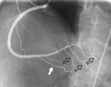
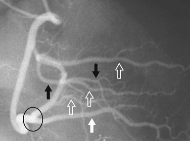
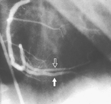
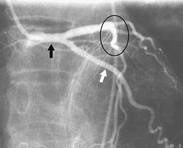
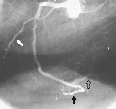
In most patients, the right coronary artery is best imaged with a combination of left lateral, cranial left anterior oblique (Cr-LAO), RAO, and Cr-RAO views. Although not used by many cardiologists, the lateral view (Fig. 7-1) has several advantages over a standard 60-degree LAO view because it usually allows better visualization of the mid right coronary artery and collaterals to the left anterior descending artery (LAD). The Cr-LAO view is necessary to lay out the posterior descending artery and posterolateral branches arising from the right coronary artery beyond the posterior descending artery (Fig. 7-2). In patients with large posterolateral branches, the Cr-RAO view projects the posterior descending artery and posterolateral branches clear of each other (Fig. 7-3). Usually, one should not use the Cr-RAO view to the exclusion of the RAO view (Fig. 7-4); however, because the body of the right coronary artery is foreshortened in the Cr-RAO view.


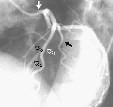
In the average patient, the left main coronary artery is projected at true length in either a posteroanterior or a shallow LAO view (Fig. 7-5). The bifurcation and proximal portions of the LAD and left circumflex artery are usually best seen in the Cr-LAO1 (Fig. 7-6) and RAO projections. Enough LAO rotation should be used to place the distal LAD between the spine and the diaphragm. In some individuals, the left main coronary artery has an up-going course. This requires using caudal angulation to eliminate foreshortening. The caudal LAO2 view is the only angiographic view that should be performed with the patient holding his or her breath in expiration (Fig. 7-7). If the patient inspires, the effect of the caudal angulation is negated; this is most common in large patients and requires more exposure than nonangulated views resulting in a degraded image. Because the distal LAD frequently is superimposed on the diaphragm in the Cr-LAO view, a lateral view is often useful (Fig. 7-8).


Dec 26, 2015 | Posted by in CARDIOVASCULAR IMAGING | Comments Off on Diagnostic Coronary Angiography

Full access? Get Clinical Tree

Get Clinical Tree app for offline access