MBT shunt
RVPA shunt
Stage I
30
20
Stage II
29
14
Stage III
32
3
Total
91
37
Age at Norwood
7.1 ± 6.7 days
5.3 ± 2.8 days
Datasets were acquired at 2 congenital cardiac centres; MBT from Evelina London Children’s Hospital, UK (ethical approval 08/H0810/058), while RVPA patients’ data were provided by Boston Children’s Hospital, USA (ethical approval IRB-P00012488). Datasets consist of balanced steady state free precession (bSSFP) cine imaging in short axis orientations acquired on a 1.5-T Philips Achieva MRI scanner according to recent guidelines [4]. Images were acquired with pixel size varying from 0.85 to 1.4 mm in the short axis plane and from 5 to 10 mm inter-slice separation. Typically, the myocardial occupies from 6 to 13 slices depending on patient’s heart size and inter-slice gap. CMR scans were acquired after Stage I at age 70 to 160 days, after Stage II at 20 to 35 months of age, and after Stage III at 3 to 14 years of age. Babies were scanned under anaesthetic.
2.2 Image Segmentation and Mesh Personalization
The systemic RV was assumed to have an elliptical shape, similarly to a healthy left ventricle (LV), and therefore it includes the septal wall. RV myocardium of all short-axis cine stacks was manually contoured by an expert at end-diastole. Trabeculations were excluded from the segmentation.
Segmented myocardial anatomies were fitted to a template mesh formed by cubic Hermite bricks [17] using the methods described in [8, 9]. The mesh fitting algorithm uses all slices with anatomical information of the systemic RV. Truncation of the basal ventricular anatomy was made at the image acquisition set, when the expert operator decides the plane that captures the most basal slice of the ventricles. A limited flexibility is provided to the template by means of the allowed degrees of freedom of the template. This results in a regularized fitting of the manual segmentation, reducing segmentation errors from human operation or from its discretized nature. All 128 scans were personalized reaching an average distance no larger than 1.5 mm to the manual segmentation. After the fitting step, the surfaces of the Hermite elements were tessellated into a triangular mesh. As a result of this process, the systemic RV anatomy was described by a set of 2834 descriptive points and each of these points is assumed to be in anatomical correspondence among all anatomies.
Differences in postural information were removed by pre-aligning all cases: all geometries were rotated to set the apical-base direction in the vertical Z axis and the right to left direction (identified by the centre of the LV blood pool manually located) in the horizontal X axis. Translational offset was removed by aligning the centre of RV blood pool. Finally, size is normalized by uniform scaling such that all segmented myocardia have a volume equal to the geometric mean of the original volumes. Notice that we are including geometries of hearts of ages from 3 months to 14 years old and without the volume normalization the most important geometrical feature results to be the size. An alternative size normalization could be to consider clinical information such as subject’s weight, height or age as was done in [11]. Nevertheless, in a preliminary study, we found that size is not a relevant remodelling pattern in this population. This also explains why traditional shape indexes (volume, mass) have not been able to identify differences between surgery procedures.
Even if CMR scans are from the same patient at different stages, the analyses performed in this work considered all the data independent as in a transversal study.
2.3 Statistical Description of the Data
A mean geometry was constructed from the 128 geometrical descriptors by averaging along the population the spatial coordinates of each descriptive point. This mean geometry (also called anatomical atlas) comprises the geometrical characteristics shared among all patients, see Fig. 1.
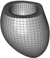

Fig. 1.
Anatomical atlas (average shape of 128 cases) of the systemic right ventricle in HLHS. Shape coordinates characterise the differences in anatomy with respect to this mean shape.
The residual geometrical information between the patients and the atlas is statistically analysed by means of principal components analysis (PCA) technique [6]. PCA performs an orthogonal linear transformation formed by the sequence of the most expressive features [14]. It is important to note that in the PCA space, the more coordinates are considered to reconstruct the data, the more accurate the reconstruction is. Figure 2 shows reconstruction errors among 128 descriptors and among the 2834 descriptive points. It can be noted that 127 PCA coordinates are required to represent the whole population (instead of the 2834 × 3 coordinates required in the Cartesian description). By considering only 16 PCA coordinates, all reconstructed instances are within 1.5 mm of accuracy (same as the mesh personalization process described before). The truncation of PCA coordinates can be also considered as a filtering of low-SNR additive noise in the data [12]. Figure 3 illustrates these concepts through a gradual reconstruction of a contour with increasing number of PCA components.
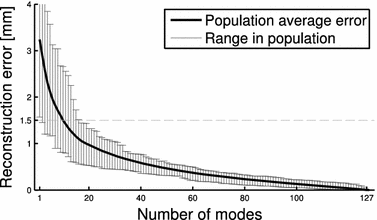
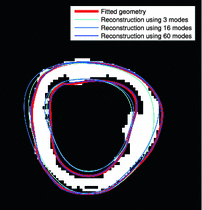

Fig. 2.
Reconstruction error in terms of the number of PCA components. 16 PCA components are enough to reconstruct all instances within 1.5 mm of accuracy.

Fig. 3.
Illustration of the mesh fitting and PCA reconstruction accuracy. It compares a segmented scan, subject to user and discretization/quantization/pixilation errors; the fitted geometry, stated to the regularization given in the fitting process; and corresponding reconstructed instances by taking into account different number of PCA components.
3 Qualitative Inspection and Quantitative Analysis
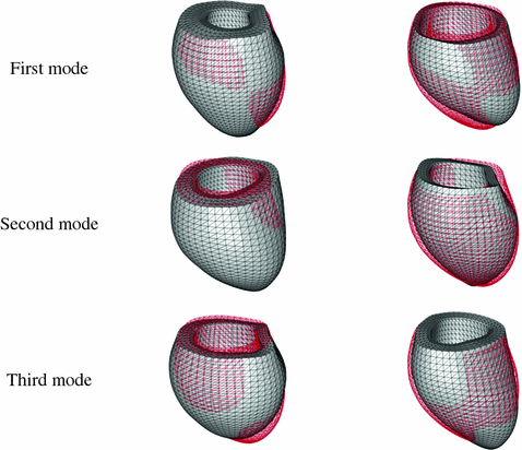
The principal modes in shape variation of the HLHS cohort, obtained by PCA, are illustrated in Fig. 4.
