Chapter Outline
Introduction
The principal structures of interest in the anterior elbow are the biceps and brachialis tendons. The biceps tendons form from its two muscle bellies and each inserts, close to one another, on the radial tuberosity. The two muscle heads can easily be distinguished from one another but the tendons are grouped close together, although the two components become more separate as the tendon approaches its insertion.
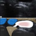
| It is important to identify both the biceps and brachialis tendons in the antecubital fossa as, if only a single tendon is present, it means that the other, usually the biceps, is ruptured ( Fig. 8.1 ). |

Biceps Tendinopathy
Biceps tendinopathy is most often due to misuse injury, although frequently there is a background predisposition. Such conditions include diabetes, systemic arthropathy, renal disease and occasionally drug use. The patient presents with antecubital pain and not infrequently a palpable mass, representing a combination of tendon and sheath enlargement.
Occasionally the mass predominates and some patients with chronic biceps tendinopathy are suspected of having a tumour.
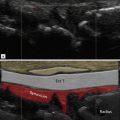
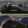
In the earliest stages, fluid gathers within the tendon sheath and synovial thickening occurs. The tendon remains normal initially with a typical reticular reflective pattern best appreciated on axial images, but also when good long-axis images are obtained ( Fig. 8.2 ). Ultimately internal signal changes representing focal tendon degeneration lead to delamination and partial tears ( Fig. 8.3 ). A hypertrophic pattern is more usual and the combination of tendon hypertrophy, fibre disorganization, fluid distension of the tendon sheath and increased Doppler signal is typical.

Related posts:


Stay updated, free articles. Join our Telegram channel

Full access? Get Clinical Tree


