30 Dorsal Arteries of the Foot
T. Rodt, M. Lee
The anterior tibial artery ends as the dorsal artery of the foot, which branches into the lateral tarsal artery and the medial tarsal arteries, and ends as the arcuate artery. This forms the dorsal metatarsal arteries, which continue as the dorsal digital arteries. In the first interosseous space, the prominent deep plantar branch is connected with the plantar arch. The other dorsal metatarsal arteries are also connected with the plantar arteries. These perforating branches can replace the dorsal metatarsal arteries.
30.1 Origin of the Arteries on the Dorsal Side of the Foot
The great variety of combinations of a more dorsal (D) or plantar (P) origin of the arteries on the dorsal side of the foot is shown. The figures do not include all the possibilities, but those omitted account for only 3 to 4% of all cases.1–5

Fig. 30.1 DDDD “normal” (20%). Schematic.

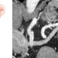
Fig. 30.2 DDDP (6%). Schematic (a), lateral oblique angiography (b), and DSA (c). Dorsal supply by anterior tibial artery (yellow marks), plantar supply (red arrow) by posterior tibial artery (red lines) and communicating branch (green arrow).
Related posts:


Stay updated, free articles. Join our Telegram channel

Full access? Get Clinical Tree


