Dual Source CT
z-Flying Focal Spot
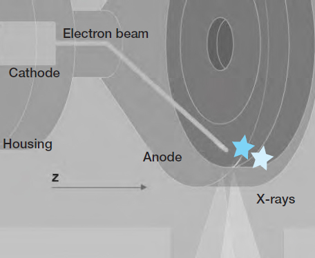
Several techniques have been established that can provide submillimeter resolution with short breath holds, even in routine protocols. The entire x-ray tube rotates in an oil bath, so that the anode is in direct contact with the cooling oil. The central cathode rotates as well. In recent years, further improvements in spatial and temporal resolution have been achieved by adding periodic motion of the focal spot in the longitudinal direction (z-axis).
Permanent electromagnetic deflection of the electron beam is used to control the shape and position of the focal spot. In this way the focal spot is made to “fly” back and forth between two different positions on the anode (indicated by the two asterisks in Fig. 198.1 ). Since the anode plate is typically angled by approximately 7-9°, this deflection translates into motion of the x-rays in both the z-axis direction and the radial direction.
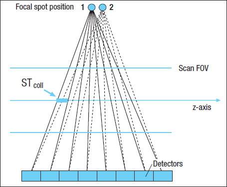
Applied to a 64-slice CT scanner, this technology can double the number of simultaneously acquired slices, say, to 64 overlapping 0.6-mm slices per rotation, similar to the sampling scheme of a 64 x 0.3-mm detector ( Fig. 198.2 ). The periodic motion of the focal spot on the z-axis can achieve a resolution equal to just half the collimated slice thickness at the isocenter (STcoll/2). This double-z sampling is no longer optimal at greater distances from the isocenter, however, and so ultimately the highest z-axis resolution that can be achieved is approximately 0.33–0.36 mm (14–15 lp / cm [47, 48].
The focal spot can also be deflected within the axial scan plane (for clarity, not shown here) to improve in-plane resolution. When this deflection is added to the scan, the focal spot flies within a roughly rectangular area that is constantly controlled by the electromagnetic deflection of the electron beam.
Data Acquisition System
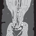
The dual source CT technique was introduced onto the market by Siemens Healthineers. These systems, called Somatom Definition, Flash, Drive and Force, incorporate two detector systems: one with a 50-cm FOV and almost 88,000 detector elements, and a smaller array. It also has two separate x-ray tubes, which can be operated in different modes. Most routine examinations can be performed with only one tube/detector pair. But cardiac patients, trauma patients, and very obese patients can be scanned at a tube output of 2 x 80 kW. This dual power mode combines the output of two x-ray tubes to obtain high performance and /or high scan speeds when specifically needed, as in CT angiography of the iliofemoral, pulmonary, and abdominal vessels (see pp. 186–188) or the coronary arteries (see pp. 184–185).
Another advantage of these new scanner systems is their speed: The shorter gantry rotation time up to 0.25 seconds can provide temporal resolutions as high as 62 ms with ECG-gated reconstruction. This feature is particularly useful in cardiac examinations at a high pulse rate, as it can reduce the number of patients who would otherwise require pharmacologic reduction of their heart rate. This new generation of scanners can completely cover the chest (approximately 35 cm) with submillimeter resolution in approximately 1 second. As a result, this innovative technology can significantly shorten the usual breath-hold times while reducing respiratory artifacts.
Detector Design
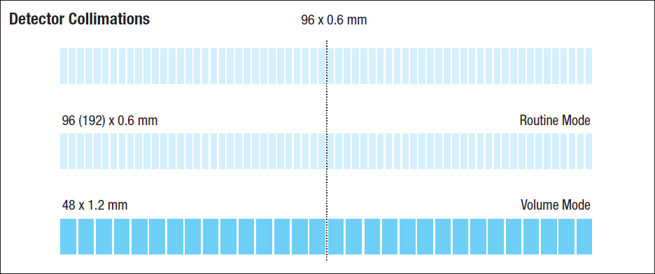
The principle of adaptive detector design was previously described on pages 10 and 11. At the time of this writing, Dual Source systems manufactured by Siemens employ a 96-row detector, which can also be used as 192-row detector (with application of z-flying focal spot, cf. p. 198). It has a collimated z-axis slice width of 0.6 mm each, resulting in a total z-axis coverage of approximately 58 mm ( Fig. 199.1 ).
As described on the previous page, the amplitude of the periodic z-axis motion of the focal spot is adjusted in such a way that two subsequent data acquisitions are shifted by one-half the collimated slice thickness along the z-axis.
Related posts:


Stay updated, free articles. Join our Telegram channel

Full access? Get Clinical Tree


