Duodenitis

 Upper GI, CECT


• Best diagnostic clue
image Upper GI: Aphthous ulcers, fold thickening, luminal spasm of duodenal bulb (± gastric antrum)

image CECT: Mural thickening of duodenum ± adjacent inflammation


TOP DIFFERENTIAL DIAGNOSES


• Cholecystitis

• Pancreatitis

• Ureteral colic


PATHOLOGY


• Gastritis commonly coexists with duodenitis
image Similar findings of mucosal erosions, fold thickening, luminal spasm


CLINICAL ISSUES


• Most common signs/symptoms
image Abdominal pain, nausea, vomiting

• Other signs/symptoms
image Gastrointestinal bleeding with deeper ulceration

• Epidemiology
image Helicobacter pylori infection and NSAID use

• Treated with proton-pump inhibitors (plus antibiotics for H. pylori)


DIAGNOSTIC CHECKLIST


• Duodenitis often coexists with gastritis

• Symptoms are indistinguishable from peptic ulcers
image Presence of only superficial (aphthous) erosions and fold thickening distinguishes duodenitis from duodenal ulcer

• Diagnosis usually established by endoscopy

image
(Left) Spot film from an upper GI series shows aphthous ulcers image 
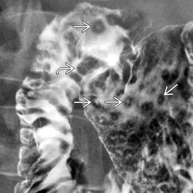
in the gastric antrum and duodenal bulb, along with thickened duodenal folds image, classic features of duodenitis and gastritis.


image
(Right) Spot film from an upper GI series shows nodular fold thickening image 
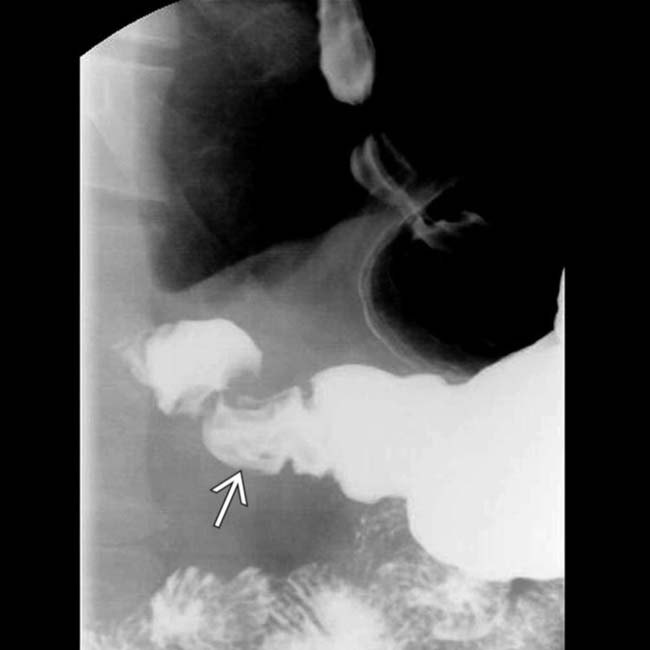
and lack of distensibility in the gastric antrum due to gastritis.

image
(Left) Another spot film from the upper GI series shows spasm and fold thickening of the duodenum image
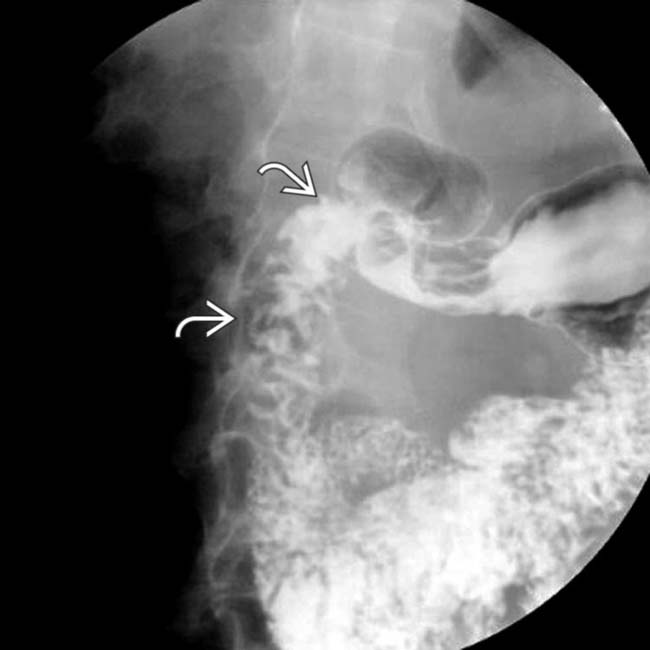
, due to duodenitis.
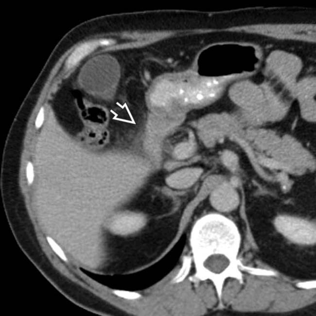

image
(Right) Axial CECT in the same patient shows luminal narrowing and mural thickening of the 2nd portion of duodenum, with surrounding inflammation image due to duodenitis.


TERMINOLOGY


Definitions


• Duodenal inflammation from any cause


IMAGING


General Features


• Best diagnostic clue
image Upper GI: Aphthous ulcers in duodenal bulb; fold thickening in antrum and duodenal bulb

image CECT: Mural thickening of duodenum ± adjacent inflammation

• Location
image Duodenum

• Size
image Ulcers 3-7 mm

• Morphology
image Discrete erosions with surrounding mound of edema in ring-like fashion


Imaging Recommendations


• Best imaging tool
image Upper GI, CECT

• Protocol advice
image Oral and IV contrast

Nov 16, 2016 | Posted by in GASTROINTESTINAL IMAGING | Comments Off on Duodenitis

Full access? Get Clinical Tree

Get Clinical Tree app for offline access