Jean Noel Buy1 and Michel Ghossain2
(1)
Service Radiologie, Hopital Hotel-Dieu, Paris, France
(2)
Department of Radiology, Hotel Dieu de France, Beirut, Lebanon
26.1 Embryology
26.2 Anatomy [, ]
26.2.1 The Cervix
26.2.4 Nerves
26.2.5 Uterine Support
26.3 Histology
26.4.1 Epithelium
26.4.2 Stroma
26.5.1 Epithelium
26.5.2 Stroma
Abstract
The cervix is separated from the uterus body by the isthmus. It is classically divided in portio vaginalis (ectocervix or exocervix), i.e., the portion that protrudes into the vagina and a supravaginal portion. It is about 3.0 cm long and 2.5 cm in diameter. The endocervical canal is limited below by the external os and upward by the internal os.
26.2 Anatomy [1, 2]
26.2.1 The Cervix
The cervix is separated from the uterus body by the isthmus. It is about 3.0 cm long and 2.5 cm in diameter.
The cervix is divided into two portions:
(a)
A portion that protrudes into the vagina called the portio vaginalis also referred to as the exocervix. This part of the cervix forms with the vaginal wall grooves called vaginal fornices (anterior, lateral, posterior).
(b)
A portion above the vagina called the supravaginal portion also referred to as the endocervix.
The portion may be divided into anterior and posterior lips, of which the anterior is shorter and projects lower than the posterior lip.
The upper end communicates with the isthmus by an upper aperture [1]. During pregnancy and childbirth, the part of the cervix closest to the body of the uterus is called by the ob/gyn the internal os. The lower end opens into the vagina by a lower aperture called the external os. The external os is connected to the isthmus by the cervical canal.
There are two longitudinal ridges, one each on its anterior and posterior walls, that give off palmate folds. They ascend laterally like the branches of a tree. The folds interdigitate to close the canal.
26.2.2 Relations with the Neighbouring Structures
(a)
Anteriorly the cervix is devoid of peritoneum. Its upper limit is in relation with the vesicouterine pouch. The supravaginal part is separated from the bladder by cellular connective tissue, called the vesicouterine septum [1] (Fig. 26.1). This septum is an anterior portion of the parametrium, which is in continuity laterally with the Mackenrodt’s ligament.
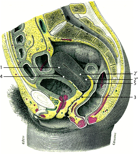
Fig. 26.1
Sagittal section through the pelvis: 1 body of the uterus, 2 cervix: 2¢ supravaginalis portion, 2″ infravaginalis portion, 3 vagina, 4 vesicouterine pouch, 5 Douglas (From Testut [1])
(b)
Posteriorly the peritoneum covers the upper part of the cervix, then the posterior fornix, then reflects on the anterior wall of the rectum to form the Douglas cul-de-sac (Fig. 26.1). On the upper part of the posterior surface of the cervix is a small transverse protrusion, the torus uterinus. On each side of the torus, uterosacral ligaments go backward on the lateral faces of the rectum to join the anterior face of the sacrum (see Chap. 17). They limit laterally with the rectum the lateral parts of the Douglas (Fig. 26.2
 Ovarian Infertility
 Premalignant and Malignant Tumors of the Vulva
 Benign Diseases of the Cervix
 Anatomy and Radioanatomy of the Pelvis and the Perineum and Anatomical Locations of Pelvic and Perineal Masses
 Congenital Abnormalities of the Mullerian Ducts
 Benign and Malignant Mesenchymal Tumors of the Uterus


Related posts:
Ovarian Infertility
Premalignant and Malignant Tumors of the Vulva
Benign Diseases of the Cervix
Anatomy and Radioanatomy of the Pelvis and the Perineum and Anatomical Locations of Pelvic and Perineal Masses
Congenital Abnormalities of the Mullerian Ducts
Benign and Malignant Mesenchymal Tumors of the Uterus
Stay updated, free articles. Join our Telegram channel
Full access? Get Clinical Tree
